DIATOMS!! JEWELS OF THE SEA 

National Diatomaceous Earth Day 

Part 2 – The Sequel

January 2, 2026

While Diatomaceous Earth (DE) may sound familiar to you (if not, read all about this chalky, gritty powder in my Part 1 – Diatomaceous Earth post), did you know it’s a product of millions of years old, long-deceased and fossilized Diatoms? Whether or not you’ve heard of diatoms, prepare to be wowed by these tiny little, microscopic ecological marvels!  Diatoms may be the primary ingredient found in DE, but as living organisms they are so much more.

Having discovered a bounty of eye-popping information about and related to diatoms, I thought it would be best presented in digestible doses. So throughout this month, diatom posts will address some of the things I wondered about and explored when studying living diatoms, the “Jewels of the Sea!”

In this post, Part 2, I’ll discuss and/or try to answer the following questions:  

What are diatoms …. plant, animal or mineral? 
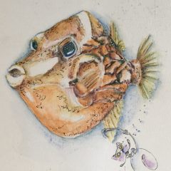
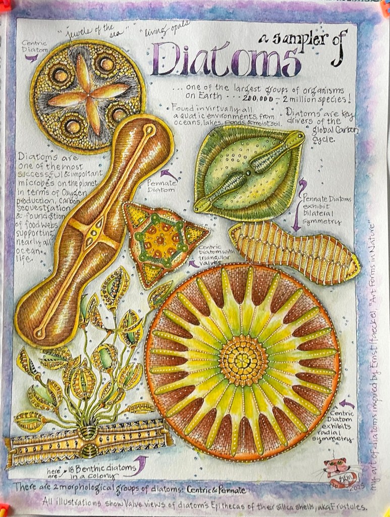
Why the nicknames, Jewels of the Sea” and “Living Opals?”  

What do they look like? 

How big are diatoms? 

Where and how do they live? 

How many species of diatoms are there?

So come take a deep dive, if you dare, on my continuing journey into the fascinating world of living Diatoms!

What are Diatoms …. Plant, Animal or Mineral?

“… a riddle wrapped in a mystery inside an enigma…” 

Winston Churchill used this phrase in an October 1939 radio speech to describe something difficult to comprehend. He could’ve been referring to diatoms. 

Diatoms are the very definition of an “enigma.” How can algae, be anything other than a plant? How can algae with obvious plant-like features also exhibit animal-like characteristics?  How can one of the most abundant minerals in the Earth’s crust, silica, be sculpted into elegant protective shells (frustules) surrounding these algae? These Diatoms? How is it possible diatoms, single-celled microscopic algae, are neither plant nor animal nor mineral, but are perfectly content and even thrive sharing biochemical features of all three?

Official Catalog of Life showing where diatoms fit in the hierarchy.

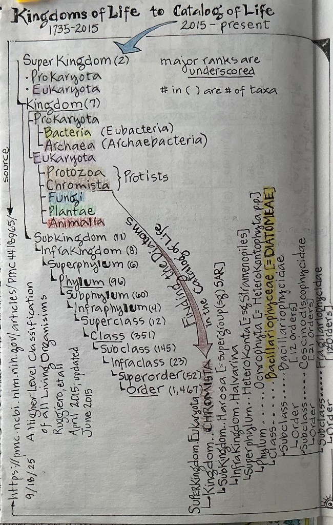
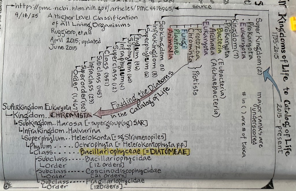
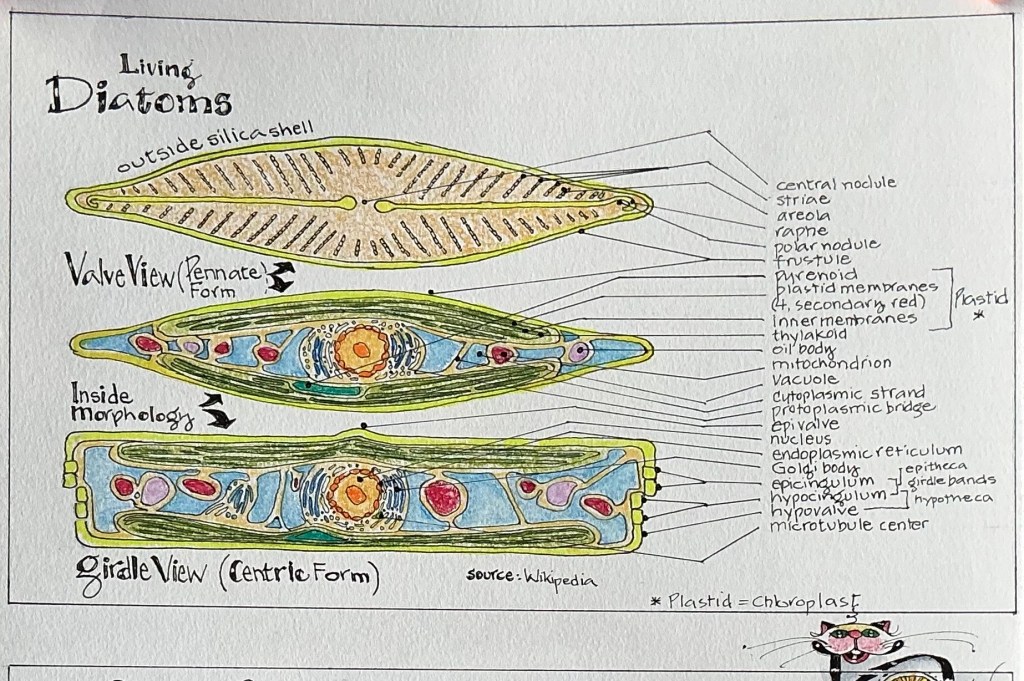
But my months-long look into living diatoms convinced me these single-celled microscopic algae (a microalgae) have earned their place as members in the Super Kingdom Eukaryota, along with animals, plants, fungi, the protists (such as algae [like seaweed; here’s where diatoms fit], amoebas and slime molds). Eukaryotes, unicellular or multicellular organisms, are considered complex when it comes to the Protoplast, the living contents of their cell(s). The Protoplast includes the cell membrane, cytoplasm, a large central vacuole, and membrane-bound organelles like the nucleus, mitochondria, Golgi bodies, one or more chloroplasts (in plants), and specialized structures like the silica deposition vesicle (where a diatom’s silica shell [frustule] is built).  The Eukaryotes represent a major group of life on Earth. 

So how did such a vast group of organisms, the diatoms, end up being partly plant-like and animal-like, as well as mineral-like? It’s about evolution …..

….. Diatom Evolution

Scientists have analyzed the genes and the proteins they encode, confirming that diatoms have had a complex history. Like other early microbes, they apparently acquired new genes by engulfing microbial neighbors. Perhaps the most significant acquisition was an algal cell, which provided the diatom with chloroplasts and the ability to photosynthesize. It’s been hypothesized that diatom ancestors branched off from an ancestral microbe with a nucleus, from which plants and animals later arose. As diatoms, plants, and animals evolved, each must have shed different genes from this common ancestor. As a result, diatoms were left with what looks like a mix of plant and animal DNA.

Official Catalog of Life showing where diatoms fit in the hierarchy.

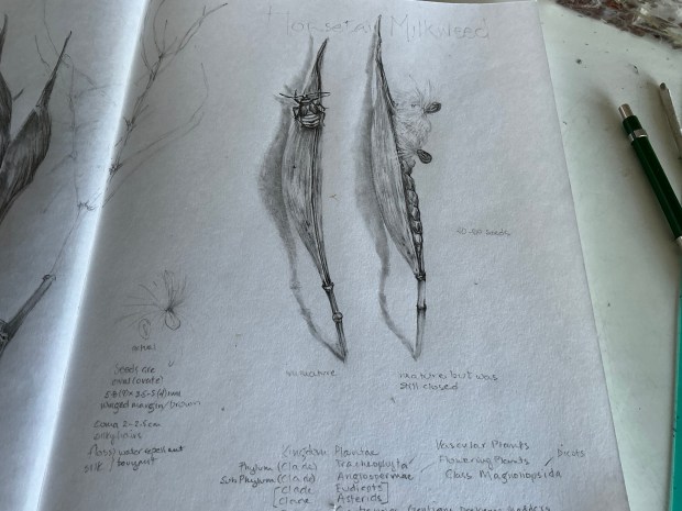
But what about that unique, silica-based frustule; a crucial part of the puzzle that so effectively protects the diatoms’ internal components while it’s alive, and plays an equally important role upon the death of all diatoms?………

….. Diatom Evolution: phase 2

Diatoms didn’t “learn” to synthesize silica but evolved a complex, genetically controlled biological process called biosilicification. This process uses specific proteins (silaffins and polyamines) to uptake dissolved silicic acid from water and precisely assemble it into intricate, ornate silica shells (frustules) within the Silica Deposition Vesicle, a feat of natural nanotechnology. This allowed diatoms to master frustule construction, making them successful in aquatic environments. 

It’s true that diatoms are:

  • a little bit “plant.” They have pigments in their chlorophyll that capture and convert sunlight into energy (sugars) through the process of photosynthesis. They use the genetically-controlled process of biosilicification to grow and assemble their unique and intricate silica frustules, similar to the way other plants, sponges, (and some animals and bacteria) do to create hard shells and/or skeletons for structural rigidity and protection. And they form the base of aquatic food chains by converting carbon into organic matter. 
  • a little bit “animal.” Some diatoms are motile, able to move (glide) using a slit-like structure called a raphe, while others use internal oil/lipid vacuoles to control buoyancy for vertical movement/migration. In addition to creating energy through photosynthesis, some diatoms also “eat” to supplement their energy and nutrients by absorbing organic matter or engulfing prey. And while all diatoms reproduce asexually, they also undergo sexual reproduction to grow, creating larger cells.
  • a little bit “mineral.” To make their unique biogenic silica frustules, diatoms must begin with the mineral silicon dioxide (SiO2), and add water. This amorphous (non-crystalline) hydrated silicon dioxide is the same material that forms opal!

And that is why diatoms are nicknamed “Jewels of the Sea” and “Living Opals!”  

The very elegant, intricately patterned, transparent frustules defracts sunlight off all the microscopic silica spheres that went into the “construction” of their glass houses, just like they were formed from priceless jewels! Just like they are “Living Opals!” 

An assortment of diatoms, centric and pennate forms (Pixabay)

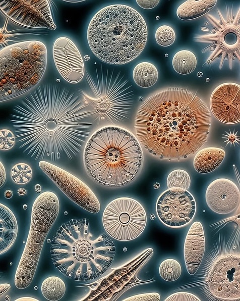
What Do Diatoms Look Like? 

First Impressions

Diatoms look like shimmering gold gems in sunlight! That’s because they live in houses literally made of transparent glass! They’re the only organism known to have cell walls composed almost entirely of amorphous (hydrated non-crystalline) silica, the natural form of silicon dioxide (SiO2).  

The glassy houses (frustules) of all diatoms are etched with intricate and beautifully-detailed patterns which aid in their identification. Each species of diatom creates frustules with unique designs, visible on both the upper (epitheca) and lower (hypotheca) surfaces (called valves) of their houses, This genetic characteristic makes one species readily distinguished from all others, by design patterns.

But these living frustules are not made of pure silica. They also have an organic layer. Their primary component is biogenic silica, a hydrated and polymerized silicic acid compound, similar to the gemstone opal. The biogenic silica, formed within the cell of the diatom in a specialized structure called the Silica Deposition Vesicle, is associated with several organic molecules such as proteins (silaffins and silacidins), long-chain polyamines, carbohydrates, and glycoproteins. 

The biogenic silica gives the frustule its rigid, intricate, and highly porous structure. The organic matrix is crucial for controlling the precise nanoscale patterning and morphogenesis of the silica structure.

Upon Closer Inspection …..

….. you’d probably notice the two Valves (the epitheca and the hypotheca) of the frustule are connected by narrow bands. These silica structures are called the Girdle Bands (or the cingulum). The girdle bands are visible when viewing a diatoms in profile; the Girdle View.

….. you’d also notice the colors of the diatoms. Regardless of the design patterning of the frustules, all diatoms appear to be either yellow-brown to golden-brown, or sometimes greenish in color. There’s at least two reasons for this: 1) because of the photosynthetic pigments found in the Chloroplasts. The primary pigment fucoxanthin (a carotenoid), masks most of the greenish hues of both chlorophylls a & c (present in smaller amounts), giving diatoms their characteristic colors or hues; and 2) the transparency of the silica frustule allows these hues to show through the internal cell wall and the frustule.

….. you’d probably notice the many and varied shapes of diatoms, with two basic forms repeated over and over again; roundish (more or less) and oblong-ish.  Yes, there are two basic forms (morphological groups) of diatoms, Centric and Pennate. Centric diatoms are usually round, have radial symmetry and are often planktonic, while pennate ones are elongated or boat-shaped, bilaterally symmetrical, and often benthic. Within both of these forms of diatoms exist a seemingly unlimited diversity of shapes, but you’d always find they are either centric or pennate. By the way, most diatoms fall within the pennate form.

Diatom colony (Pixabay)

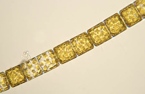
….. you’d probably notice there are diatoms that appear to be stuck to a lot of other diatoms. These are the ones that live in Colonies, from the photic zone down to mid- and deep ocean levels, and may be attached to substrates (like corals or sediments) or free-floating in the water column. Colonies can be either centric or pennate with some forming colonies in chains or  zigzags.

and perhaps lastly …..

….. you’d probably notice the pores on the frustules of all diatoms, and slits that are present some.  Those tiny Pores allow for nutrient uptake and waste removal, while only some species have slits called Raphes, which allow the diatom limited movements.

How Big Are Diatoms?

Almost all known diatom species are microscopic. To comprehend just how tiny these single-celled organisms actually are, it’s helpful to compare them to another tiny and common creature; one that’s clearly visible with the naked eye; the Ant. 

But first, a bit about Units of Measure at the Microscopic and Macroscopic Scales.

Things that are Microscopic, like pollen grains, bacteria, dust, filter ratings, and light wavelengths, are measured in microns or micrometers, two names for the same unit of length. “Micron” is the older, more common term, while “micrometer” is the official International System (SI) World’s standard name for this unit of measurement. Both names are abbreviated as “µm.”

One micrometer is equal to one-millionth of a meter (m) [also represented as 10-6 ] or one-thousandth of a millimeter (0.001 mm).

Things that are Macroscopic, such as people, reptiles, coins, trees, rocks, mountains, and everyday objects, exist at a scale much larger than a virus. Macroscopic things can be seen, measured and observed with the naked eye, without needing magnification, and are measured using the units of the SI system/the metric or base-10 system; or the U.S. Customary (non-base-10) system. 

Today, the World’s standard or SI system is the most widely adopted for its coherence and simplicity. With that in mind, 1 mm = 1000 μm. 

And one last thought regarding scale ….. a macroscopic view shows the whole thing (e.g., a piece of metal or a body of water), while a microscopic view reveals their atoms and molecules.

A common ant on moss …. The moss makes a perfect habitat for microscopic diatoms (Pixabay).

Now on to Diatoms vs Ants – Size Ranges

Because diatoms are almost entirely microscopic, they are measured in micrometers. These single-celled organisms range in size from a minimal dimension of less than 1 micrometer (μm) (0.001 mm or 0.00003937 inch) to a maximum of over 5,000 pastedGraphic.pngμm (5 mm or 0.2 inch). However, the typical size of most diatoms is generally 20 to 200 pastedGraphic.pngμm (0.02 to 0.2 mm or 0.0008 to 0.008 inch). 

Because ants are entirely Macroscopic, they are measured in millimeters. Ants range in length from about 0.75 to over 52 mm (0.03 to 2 inches).  But the length of most common species vary from 1.5 to 13 mm (0.06 to 0.5 inches). 

Diatoms and Ants – Relative Comparison 

Comparing the smallest ant (0.75 mm) to one of the largest diatoms (5 mm), the diatom is actually larger. However, the vast majority of diatoms are much smaller, with the smallest diatoms less than 1μm (0.001 mm), making them about 750 times smaller than the smallest Ant. 

But if you compared a typical diatom (0.1 mm long) to a typical ant (7 mm long), an ant is 70 times longer than a diatom.

Basically, diatoms, which can appear as a speck of dust to the naked eye, exist on a Microscopic scale, while ants are Macroscopic and belong to an entirely different order of magnitude in size. 

Where and How Do Diatoms Live? 

Diatoms, found worldwide, are primarily Aquatic in nature and live their lives entirely in aquatic habitats. But many Terrestrial species have adapted to live very well in non-aquatic, moist habitats. 

Aquatic Diatoms

Those species dependent on aquatic habitats exist in cold Rocky Mountain streams to hot thermal springs in Arkansas; from polluted pools to road side ditches; from ponds and lakes to streams, rivers, seas and the oceans. They thrive as free-floating (plankton or phytoplankton), attached, or bottom sediment-dwelling (benthic) organisms. For the most part, Aquatic Diatoms live in the sunlit upper layers (photic zone which goes 200 meters or 656 feet deep) of all waters, from oceans to freshwater lakes. 

Terrestrial Diatoms

In moist or damp conditions, where moisture levels fluctuate or are unpredictable, there are diatom species that thrive in soils, sediments, or attached to mosses, tree trunks, rock faces and even brick walls. They exist happily in ephemeral marshes, bogs, fens and swamps. Other species make their home on the feathers of diving and wading birds, and waterfowl! Some even prefer the nooks and crannies of lichens to stay humid.

Adapting to Drought

Diatoms are very resilient. To survive drying out, they form resilient films on surfaces of temporary water bodies. Many secrete a protective, glue-like mucus (mucilage) that helps them cling to surfaces and retain moisture.  To endure a lengthy drought when conditions become too dry, they transition into a low-energy resting stage, conserving resources until water returns, a process sometimes involving nitrate respiration. So, while diatoms need water to grow and reproduce, certain diatom species are masters of surviving desiccation in non-aquatic, moist environments. 

When All Else Fails …..

Then there those diatoms, the endosymbionts, that live inside other small organisms, such as dinoflagellates and foraminifera. Both of these organisms are types of plankton, specifically phytoplanktons (microscopic, single-celled organisms, free-floating (drifting) and living in the photic zone of aquatic habitats). Although the majority foraminifera live as benthic (deep water) organisms.

Bottom line ….. Diatoms can thrive wherever there is sunlight, water, carbon dioxide, and the nutrients necessary for their growth and survival.

How Many Species of Diatoms Are There?

One of the largest groups of organisms on Earth, there are about 25,000 species of diatoms that have been documented, to date, each with their own unique shell (frustule). With new ones being discovered every year, scientists studying diatoms2 estimate there may be as many as 200,000 to 2 million species worldwide-wide.

Because diatoms exist in large numbers in most bodies of water throughout the world, if you collected just one liter of seawater, it can contain as many as ten million individual diatoms! This makes diatoms the most abundant type of phytoplankton, with the greatest numbers existing in cold oceans.  

2 The study of diatoms, known as Phycology, is in a branch of botany concerned with seaweeds and other algae. A scientist studying Diatoms may be Protistologist, Diatom Paleontologist, Ecohydrologist, Environmental Botanist, Botanist, Diatomist, Paleolimnologist, an algal physiologist, a genomicist

or a Paleoceanographer.

It’s a Wrap! 

DIATOMS!! JEWELS OF THE SEA//Part 2 – The Sequel

…………………………………………………………………………………………………

Hope you’ve enjoyed learning some fascinating facts from this introduction to living diatoms. Spread the joy and tell all your family and friends! They will be so impressed with your new-found knowledge of diatoms, and may even want to learn more about these Jewels of the Sea!

As I was compiling and organizing my notes for Part 2, many additional questions about diatoms and their compatriots popped into my mind. I’d love to know if you came up with some as well. If you share your questions with me, and I’ll dig around for answers. Also, if you have any questions about or clarifications needed regarding the information in this Part 2 introduction, I’d welcome them via message or comment on the post. 

Meanwhile, here’s a teaser from the upcoming Part 3 in my Diatom Series. Things I’ve wondered about while researching diatoms: 

Do diatoms breathe? Eat? Reproduce? How long do they live? What good are diatoms? What do they eat and are they food? What’s their relationship with the global carbon cycle? Are diatoms all good and wonderful? Do they have a bad side?

………………………………….     ………………………………………………………….

If you feel yourself becoming a “Diatomic Geek,” you may find interest in browsing the following Glossary of terms. If a term used within the text of my post is in bold font and italicized, it is (or will be) in the Glossary. (Fair warning: my Glossary continues to be a work-in-progress. Look for an updated version in Part 3 of this series on diatoms.)

Glossary

Archaea(ns) – single-celled microorganisms similar in structure to bacteria, but evolutionarily distinct from bacteria and Eukaryotes. They are obligate anaerobes living in low oxygen environments (e.g., water, soil), and form a commensal (symbiotic) relationship in ruminant and human intestines. When Archaea is the commensal, it benefits from the relationship with the host without causing harm, or may provide certain benefits to the host (i.e. the human intestines).

Archaeans may be the only organisms that live in some of the most extreme environments on the planet; habitats hostile to all other life forms. Some live near thermal rift vents in the deep sea at temperatures well over 100 degrees Centigrade. Others live in hot springs, or in extremely alkaline or acidic waters. They thrive inside the digestive tracts of cows, termites, and marine life where they produce methane. They live in the anoxic muds of marshes and at the bottom of the ocean, and even thrive in petroleum deposits deep underground. They survive the dessicating effects of extremely saline waters.

However, archaeans are not restricted to extreme environments; they are also found, in abundance, in the plankton of the open sea.

Autotroph (aka Producers) – an organism that produces its own food using external energy sources like sunlight (through photosynthesis) or chemical compounds (through chemosynthesis).

Biogenic silica (bSi) – is amorphous, opal-like hydrated silica (SiO2 · nH2O) produced by living organisms, forming structures like diatom shells (frustules), sponge spicules, and plant phytoliths, playing roles in structural support and defense against stress. It’s a key component in marine and terrestrial ecosystems, with significant deposits like diatomaceous earth (DE) from fossilized diatoms.

Biosilicification – the biological process where living organisms take up soluble inorganic silicon (silicic acid, Si(OH)4) from their environment and convert it into solid, polymerized, insoluble silica (SiO2) to form hard structures like shells, skeletons, or cell walls, crucial for diatoms, sponges, and plants, and even occurring in some bacteria and mammals. This process is a form of biomineralization, where organic molecules are used to control the precise formation of these silica materials. 

How it works 1) the organism must uptake (absorb) silicic acid; 2) then polymerization takes place, where inside or outside the cell, the silicic acid condenses, forming chains of silica with the removal of water; and 3) specialized proteins and other macromolecules guide (control) this process, creating complex, often intricate silica structures (biogenic silica) like those found in diatom frustules.

Carbon fixation – diatoms “fix” carbon by removing inorganic carbon dioxide (CO2) from the atmosphere and converting it into organic carbon in the form of sugar (glucose). During this process, oxygen (O2) is released back into the environment, producing 20-30% of the air we breathe.

Catalog of Life – the 1998 Kingdoms of Life were replaced in 2015 with today’s adopted Catalog of Life: At the top most tier or classification rank are two Super Kingdoms, Prokaryota and Eukaryota. The next descending rank the Kingdoms, of which there are seven, (under Prokaryota are two Kingdoms: Bacteria and Archaea); under Eukaryota are five Kingdoms: Protozoa, Chromista, Plantae, Fungi, Animalia).

Centric – a diatom withValves (cell walls) that are radially symmetrical.

Chloroplast – also called a Plastid, a chloroplast is photosynthetic organelle that absorbs light molecules (sun’s energy) through chlorophyll a & c, turning it into chemical energy by way of photosynthesis.

Diatom – a microalgae that forms a significant part of the food chain in moist soils and aquatic (marine and freshwater) ecosystems. They form long-chain fatty acids that are an important source of energy-rich molecules and provide a critical food source for the entire food chain, from tiny zooplankton to fish to whales. So it can be said that diatoms feed oceans, lakes, streams, rivers and associated wetlands and riparian areas. 

Dinoflagellates – single-celled microscopic organisms with two whip-like tails (flagella), making them motile. They are found drifting in large numbers in marine plankton, specifically phytoplankton; also found in fresh water. Some produce toxins that can accumulate in shellfish that, when eaten, results in poisoning. Dinoflagellates form crucial part of the ocean’s food web, producing a significant portion of the world’s oxygen, dinoflagellates are known for causing red tides and bioluminescence.

Endosymbiont – an organism that lives inside another organism (its host), forming a symbiotic relationship, usually where both (mutualism), but sometimes one benefits at the other’s expense.

Ephemeral – something that lasts for a very short time.

Eukaryotes – organisms characterized by complex cells; a membrane-bound nucleus that contains DNA, and other membrane-bound organelles like mitochondria.  Eukaryotes can be unicellular or multicellular, fall within the Super Kingdom Eukaryota that includes animals, plants, fungi, and protists (such as algae [like diatoms and seaweed], amoebas, slime molds) The Eukaryotes represent a major group of life on Earth.

Flagella – an appendage that provides motility.

Foraminifera (forams) – a single-celled animal with a perforated chalky, calcium carbonate shell, through which extend slender protrusions of protoplasm. Most forams are marine species – some float in the water column in the photic zone (planktonic); most live on the sea floor (benthic). When benthic forams die, their shells form thick ocean-floor sediments. 

Frustule – the external, silica cell wall of a diatom. The frustule is composed of two valves and the girdle bands. The upper valve, or epitheca, is slightly larger than the lower valve, or hypotheca. The epitheca overlaps the hypotheca similar to the halves of a pill box or a Petri dish.

In diatoms, frustules are not made of pure silica. They also have an organic layer. Their primary component is referred to as biogenic silica. (Read more about the make up of biogenic silica under the definition for “Silica” below. 

Girdle bands – the bands (aka cingulum) that connect the two Valves (epitheca and hypotheca). The girdle bands are visible when viewing a diatom in profile. (Girdle = Copulae)

Girdle view – the profile view of a diatom; view appears rectangular in both Centric and Pennate diatoms.

Kingdoms of Life – in 1969, the five major groups of life – aka Kingdoms of Life – were classified as: 1) Fungi, 2) Animalia, 3) Plantae, 4) Protista (the water molds, brown algae and diatoms); and 5) Monera (the bacteria and archaea). In 1990, there were three Domains: 1) Eucarya (including diatoms), 2) Bacteria, and 3) Archaea. Another change was made in 1998, now with two Empires (Prokaryota and Eukaryota) that ranked over six Kingdoms (Prokaryota ranked over one Kingdom, Bacteria; Eukaryota ranked over five Kingdoms: 1) Protozoa, 2) Chromista, 3) Plantae, 4) Fungi, and 5) Animalia.)

Pennate – a diatoms with Valves (cell walls) that are bilaterally symmetrical. 

Plastid – also called a Chloroplast, a plastid is photosynthetic organelle that absorbs light molecules (sun’s energy) through chlorophyll a & c, turning it into chemical energy by way of photosynthesis.

Photosynthesis – the chemical process where plants and some other organisms with chlorophyll, through the use sunlight, combine inorganic carbon dioxide with water to form carbohydrates (sugars), releasing oxygen as a byproduct.

Prokaryotes – a microscopic single-celled organism without organized internal structures; that has neither a distinct nucleus with a membrane nor other specialized organelles. Prokaryotes include the bacteria and cyanobacteria.

Protists – a diverse group of complex Eukaryotes  (organisims with a nucleus and organelles) that don’t fit into the categories of animals, plants or fungi. Protists are diverse life forms, mostly single-celled but sometimes colonial or simple multicellular forms, found in aquatic environments. They are incredibly varied, acting like animals (amoebas), plants (algae), or fungi (slime molds), and playing vital roles in ecosystems as producers and decomposers. The Protists are split into two Kingdoms, Protozoa and Chromista, under under the Super Kingdom Eukaryota. Diatoms fall under the Kingdom Chromista as described in the Catalog of Life.

Protoplast – refers to the living cell contents of diatoms, including the cytoplasm, nucleus, and other organelles, and is contained inside of the rigid silica cell wall. The protoplast is the part of the diatom that grows and divides. During division the protoplast duplicates, splitting into two complete daughter cells. But unlike typical plant cells, before the daughter cells can separate into two complete diatoms, a new silica valve is formed for each daughter protoplast to pair with one of the parent’s valves. Now the complete daughter cells separate, each with its own new species’-unique external silica shell (the frustule).

Raphe – a slit or groove opening in the silica cell wall (frustule) of a pennate diatom to allow it gliding motility and attachment to a substrate by secreting sticky mucilage, allowing these diatoms to move along surfaces and find nutrients.

Red tides – more appropriately described as Harmful algal blooms, or HABs, occur when colonies of algae—plant-like organisms, such as dinoflagellates, that live in the sea and freshwater—grow out of control while producing toxic or harmful effects (death, in some cases) on people, fish, shellfish, marine mammals, and birds. Eating organisms affected by the toxins, like fish and shellfish, make them dangerous to eat. The toxins may also make the surrounding air difficult to breathe. One of the best known HABs in the U.S. occurs nearly every summer along Florida’s Gulf Coast. This bloom, like many HABs, is caused by microscopic algae that produce toxins that kill fish and make shellfish dangerous to eat. As the name suggests, the bloom of algae often turns the water red.

Silica –  a mineral, specifically a compound of silicon and oxygen, called silicon dioxide (SiO₂), is one of the most abundant minerals in the Earth’s crust. Silica is most commonly found as quartz, but also in other solid forms like cristobalite, tridymite, and opal.

In diatoms, the living frustules are not made of pure silica. They also have an organic layer. Their primary component is referred to as biogenic silica, a hydrated and polymerized silicic acid compound, similar to the gemstone opal. The biogenic silica, formed within the cell of the diatom in a specialized structure called the Silica Deposition Vesicle, is associated with several organic molecules such as proteins (silaffins and silacidins), long-chain polyamines, carbohydrates, and glycoproteins. 

Silica Deposition Vesicle – a specialized internal structure of a diatom, where its silica shell (frustule) is built before being exported. 

Stramenopiles – a phylum (also referred to as a clade), synonymous with the phylum Gyrista

within the Protista Kingdom. The Stramenopiles (aka Heterokonts) include Water Molds, Brown Algae (Sargassum species, Fucus species, and Kelps), and Diatoms (Centric and Pennate).

Valves – the cell walls of diatoms are made up of two Valves; top (epitheca) and bottom (hypotheca). The top Valve is slightly larger so it overlaps the bottom Valve, like a pill box or Petri dish.

Valve view – the view of a diatoms when looking face-on at one of the two Valves.

Under Siege! 

A Battle of Wits

Conflict of Interest

November 24, 2025

Were you ever so challenged by something so clever, while at the same time so frustrated with something so beautiful? No, no, wait….. that question may be more complicated than need be. Let me put it this way …… 

Were you ever at your wit’s end finding a solution to a seemingly simple problem that you thought was obviously and repeatedly staring you right in the face?

My reply? Yes!

It’s All About the Genes

Meet the Northern Flicker (Colaptes auratus) … or more specifically, the Western red-shafted flicker (C. auratus ssp. cafer)*, a gorgeously flamboyant and noisy member of the Woodpecker family, that’s common throughout its western range.**  And as woodpeckers do so well, they peck and peck and hammer and drill with the determination and force of a jackhammer*** on nearly any vertical (preferably wooden) surface. They’re single-minded, from start to finish, when it comes to creating a cozy nesting or roosting cavity, whether in a tree trunk or into your home.  (More about that in a bit.)

Flicker ID – 101

How do you know a Flicker has laid claim to your place? Well, he’s a big, heavy-bodied bird, and when flying overhead, your first thought might be “Crow!” At 12-14” long, with a wingspan of 18”-21”, the size is right. But as he flashes a large showy white rump patch bookended by reddish-orange underwings, you realize he’s not black. Anything but! As his flight slows and dips you notice his brown back is marked with narrow black bars. In preparation for landing, with wings open wide, he vertically aligns his body and feet with the wall, exposing a pale gray belly with bold black spots and a chest-wide black patch. Two strong clawed-toes up, two down (zygodactyl), and a stiff wedge-shaped tail adjusted as a brace, he taps out a few test spots, drawing your attention to his long and heavy bill, on a slate gray head broken by a buff-brown crown, a bright red whisker (male), and light gray cheeks. 

Male Western red-shafted flicker in flight. Note white rump patch (unsplash.com)

On a crisp cool Autumn morning, as you watch in horror …… 

Before you can declare, “It’s a Male Flicker!” ……

This bigger-than-life bird has landed, tested, and pecked away at his chosen spot  170 times in 10 seconds! He’s created an entry hole about 3” wide, right through the stucco and foam sub layer. This determined Flicker knows winter is coming and he intends to drill into our home, making a cozy roosting cavity in which to hunker down until Spring!  

Oh no, No, NO!

We love Flickers and have no wish to harm this beautiful bird.**** But he’s already caused enough damage (23 feet high on the wall) that needs immediate repair. So I clap my hands and holler loudly (something unintelligible), and off he flys to a nearby snag to see how serious my noise-making was.

That’s the story of Flicker hole #1 

Oh Not Again, and Again, and Again!

Since early November, our resident Flicker (I call him Jack), has continued to return many times, usually between sunrise and 10am. Sometimes he’ll make a fly-by before sunset. Often his quiet arrival escapes our notice; either we’ve been running errands, we’re out hiking with Luna, or enjoying a short roadtrip. These are the times he’s been able to drill six 3”-wide holes on the initial wall, and another 3”x6” hole just around the corner which was so deep, he almost penetrated the interior of Roy’s woodshop! This gives a whole new meaning to the term “Airbnb!”

After a few weeks up and down our fully-extended extension ladder to make a 2-step/2-day repair job/hole, we were making ZERO headway. Jack, unable to resist the need to drill him a roost cavity, was always one hole ahead of us. And because he didn’t hesitate to redrill newly repaired holes, was there something we were doing wrong?

All Flicker painting are larger than life, because that’s how they seemed to me!

It’s an Education in Biology and Patience

So we learned to listen for his noisy “kerrreee” scream-like call announcing his presence from one of Jack’s many favored perches around the house. Unless we missed it, his territorial call would put us on high alert, ready for action. We also listened for his series of warm-up test pecks that usually sounded inside the house. This “alarm” would catapult one or both of us from a comfy chair and run outside yelling and clapping our hands. 

Between listening, running, clapping and yelling (and wondering what the neighbors might be thinking), I discovered a few interesting things on-line…..

  1. Woodpeckers can’t resist drilling holes in synthetic stucco. This product provides the perfect surface for woodpeckers to hammer. When they begin tap pecking, it creates a hollow sound because the synthetic stucco includes a foam layer. The woodpeckers peck through the hard outer surface into the foam where it is easier to create a larger cavity to nest. 

#1 …. Our entire home happens to be covered with synthetic stucco! While this might explain Jack’s insatiable desire to drill his roosting cavity into our home and not into one of the surrounding hardwood piñon pines, we’re not going to replace  the stucco. 

  1. Basil, mint, cinnamon and/or lavender are suggested as natural, non-toxic deterrents for woodpeckers, who dislike strong aromas. The scent of basil, in particular, can be overwhelming and confusing to woodpeckers. Crushing one or a mix of these herbs with adding a bit of water, creates a green slurry that can be filtered and applied with a spray bottle to the affected area(s).

#2 …. This idea was worth a try, especially since there’s still have basil and mint growing in the garden. After collecting several handfuls of each, I popped the mix into the food processor with a bit of water and flipped the on switch. Gathering the resulting slurry, I filtered it through paper towels and collected the liquid for a spray bottle. That was several weeks ago, and with every hole repair, Roy’s been thoroughly soaking first the patch job then follow-up stucco coating with the basil/mint spray. It’s hard to know if it’s actually working, but the initial drilling sites haven’t been redrilled in the past week. It could also be that Jack is gone; pushed out with one of our heavy rainstorms.  Or he’s begun drilling more recent holes over the RV garage door. With each repair, Roy continues to spray the basil/mint mix.

  1. The Federal Migratory Bird Treaty Act*** provides protection for Flickers (and all woodpeckers), making it illegal to harm or kill them. But when warranted, migratory birds can be killed under a depredation permit issued by the Law Enforcement Division of the USDI-Fish and Wildlife Service (USFWS). Authorization by the relevant state wildlife agency also may be required before lethal control methods are initiated. Sound justification must be present for the issuance of depredation permits.

#3 …. Applying for a depredation permit may be our last resort, if Jack and his cohorts threaten to turn our brand new home into Swiss cheese. 

AGAIN!

That’s the story, almost. This clear Conflict of Interest; an obvious Battle of Wits, continues. Just yesterday, one of the holes Roy patched above the RV garage door was redrilled this morning!

Oh Good Grief!

 It’s already been repatched and resprayed, and while writing this story in my studio with window cracked and a clear view of the patched hole, I’m sure to hear and see that gorgeously determined Flicker if he returns to jackhammer away, once again, into a side of our home!

I’d love to know if you or anyone you know has a proven solution to this natural dilemma. Meanwhile ….. 

Thanks for stopping by, and Happy Thanksgiving!

……………………………………………………………………………………………………………

*Northern Flickers are divided into 2 subspecies, the Western red-shafted flicker (C. auratus ssp. cafer) and the Eastern yellow-shafted Flicker (C. auratus ssp. auratus). The red-shafted subspecies is found throughout Mexico, western and west-central U.S. (where it is common all year long), and British Columbia, Canada. The closely related yellow-shafted subspecies, which is highly migratory, is found in eastern and east-central U.S., the Canadian provinces and Territories (except B.C.), and far north into AK. 

**Where the range for both subspecies overlaps (in the ‘lower 48’), a lot of hybridization occurs. It’s common to see a red-shafted flicker with more orange feather shafts and/or shades of yellow-orange on the underside of their flight feathers. The same holds true for the yellow-shafted hybrid. Otherwise, appearances differ notably between both subspecies of the Northern Flicker, primarily where the malar (mustache), nape pattern (back of the head below the crown), face color, and tail and flight feathers are concerned. See the table below for non-hybrid subspecies characteristics. For hybrids, any color and pattern variation(s) and combination(s) you can imagine have probably been found! 

Northern Flicker subspeciesRed-shafted Yellow-shafted
Face colorGrayBuffy to warm, light brown
Malar colorMale: red Female: brownMale: black Female: brown
Nape color & patternGray, unpatternedMale: Red crescent on gray Female: gray, unpatterned
Feather shaft/under flight feathersPinkish to reddish to redYellow

***A woodpecker can peck wood 17x/second, and from 8,000-12,000x/day! Really! And they can drill into wood at a force 10x greater than a football tackle that would cause a concussion. On the November 17, 2025 episode of the Science Friday (SciFri) podcast, biologist Nick Antonson stated that woodpeckers can peck 20-30x their body weight. Now that’s amazing for a Flicker that weighs about 6 ounces! 

****Because we had no desire to harm the Flicker(s) drilling into our new home, even when we reached our point of extreme frustration, we wanted to ensure our deterrent efforts aligned with wildlife regulations; especially with the Federal Migratory Bird Treaty Act (Act). Flickers (and all woodpeckers) are considered a migratory non-game bird species, and protected under the Act. It’s illegal, punishable by fine and/or imprisonment, to harm or kill them. 

Male Western red-shafted woodpecker with his tail braced against the branch(bird pixels.com)

EggTober 2025 ….. the Full Four Submissions + Bonuses

November 15, 2025

It’s a wrap! EggTober 2025 – the Full Four Submissions- all in one place!

My 3 minute YouTube video (see link below) features all 32 annotated bird egg paintings submitted for Inktober2025, along with several bonuses you won’t want to miss!

1) a pair of never-seen-before annotated Common Nighthawk eggs painted for the cover of my 6”x9” handmade journal ………

2) an index, by common name and date of appearance, of the 32 bird eggs painted and annotated between October 1 and November 1, 2025 ………

3) an accordion book attached to the inside back cover that has all the fascinating details about each of the 8 layers of an avian eggshell

YouTube Video Link

For some reason, I’m unable to embed my YouTube video directly into this post. This is rather inconvenient, huh! So until I can troubleshoot the player issue, I’d love for you to click on the link below to view the video on my YouTube channel!

https://www.youtube.com/watch?v=keiOCtFwASo


And That’s All Folks!
Thanks so much for joining me on this fun educational adventure.

P.S. If for some reason you’re unable to view the video, please let me know.

EggTober 2025/Submission Four ….. Days 25 – 31

It’s All in a Day’s Work! 

November 3, 2025

EggTober 2025 has officially ended! And Wow … I’m so eggs-cited to share Submission Four, the finale, with all-y’all!  The eggs showcased for Days 25-31 +1 (yes, another random bird flew in and laid a bonus clutch on the last page) are all snug in their New Mexico nests. Also, you’ll notice a bit more text surrounding those nests.   

Following Submission Three, where my narrative focused on the different layers of the eggshell membrane, I was curious about how avian eggshells are constructed. Then I wanted to know more about the entire reproductive system of birds, from ovulation to fertilization, to egg laying. And Then I couldn’t rest without knowing about the embryo, the yolk, the egg white, and all the bits and pieces you see when cracking open an egg! I didn’t know any of this, and if you don’t know, you’re head is about to explode in wonder!

October 25 and 26

The Avian Oviduct and Egg Formation

It takes about one day to build an egg. But to prepare for the journey, about 7 to 9 days before the egg will be laid, the Yolk must be formed. Here’s the story of …..

The Yolk …..

….. formed in one of the many Follicles of the Ovary, the vitellus or Yolk begins as an immature Ovum that is stimulated to enlarge over several days by receiving deposits of yolk material. Once growth is initiated, over the next 7 to 9 days the Yolk’s formation intensifies as 99% of its nutrient-rich layers are deposited until it’s fully formed. Now mature, the Follicle ruptures and the Yolk is released in a process called Ovulation.

And this is where the Journey through the Oviduct begins! Over the next 24 hours, the developing embryo acquires the many layers it needs for nourishment, respiration, and protection until it’s formed into a perfect egg ready for laying. The entire journey takes place in the Oviduct …..

….. which can be divided into several regions: the Infundibulum, Magnum, Isthmus, Uterus, and Vagina. What happens in each of these regions is nothing short of fascinating!

The Regions of the avian oviduct

Infundibulum – After Ovulation, the Yolk immediately enters the funnel of the Infundibulum (the Ostium) where the Ovum is fertilized and the Chalazae is formed. With the help of ciliary action, it takes 30 minutes for the Ovum to move through this region of the Oviduct to the next, the Magnum

Magnum It’s this region where the Yolk, together with the now developing embryo and the Chalazae, gets a protective coating of protein-rich Albumen, a process that takes about 3 hours to complete, before moving along to the Isthmus.

Isthmus – The Inner and Outer Shell Membrane Layers and the Nucleation Sites are formed in this region of the Oviduct. This takes about an hour before the package is delivered to the Uterus

Uterus This is where five of the six layers of the outer Eggshell are formed. They are the Mammillary Layer and Mammillary Bodies, Organic Matrix Layer, Crystalline Palisade Layer, Vertical Crystal Layer, and Shell Pigment Layer. Also, as much as one-third of the protein in an egg is added while in the Uterus. 

It takes about 20 hours for the egg to move through the Uterus while these layers are deposited. And as the egg moves, it twists and turns. When it’s time to add pigment, the rate of rotation adjusts to ‘paint’ the species-specific patterns we see on the eggshell, leaving a visual record of the egg’s movement in the Uterus. For example, if streaks or elongated tracks on the shell are required, movement is more rapid than when creating round spots, blotches, speckles, or bands. After pigment is added to the outer shell structure, the layers of background color and any markings are enclosed in calcite crystals. 

Vagina – The last region of the Oviduct where the Cuticle (the Bloom) is added to the shell just before the egg is laid. 

The egg is now complete. The entire process, beginning with the release of the mature Yolk and Ovum from its Follicle until the egg’s final touches, takes 24 hours! And the timing ensures the egg is always laid sometime during the day.

………………………………………………………………………………………………………………

Glossary and Other Interesting Things

Albumen – refers to the “white” of the egg. The Albumen is an effective barrier against microbes that might cross over and cause disease in the developing embryo. Albumen contains over a hundred antimicrobial proteins.

October 27 and 28

Chalazae – two spiral bands of white, stringy tissue that suspend and hold the Yolk in the center of the Albumen.

Crystalline Palisade Layer – A  tough, mineralized structure of calcium carbonate crystals that grow into dense columnar units above the Mammillary Bodies (the mineralized base of the crystalline eggshell). The amount of shell deposited is determined by the time spent in the Uterus.

Eggshell – Depending on species, the eggshell represents from 9-15% of the total weight of the egg. 

Eggshell Formation – The Crystalline Palisade Layer is essential to the process of eggshell formation which takes place in the Uterus. 

  • Nucleation: The process begins at the Nucleation Sites, located on the External Shell Membrane, which are the specific places where the mineralization of calcium carbonate begins. 
  • Initial growth: As calcium salts are deposited and crystallize, inorganic calcium carbonate crystals grow around and outward from the Nucleation Sites, forming the cone-shaped Mammillary Bodies; the first, innermost calcified layer of the eggshell, anchored to the External Shell Membrane. This base of mineralized shell provides mechanical strength, and serves as the main source of calcium for the developing embryo’s skeleton.
  • Maturation – The calcium carbonate crystals continue to grow into the dense columnar units that form the Crystalline Palisade Layer, creating a tough, mineralized structure that protects the embryo. The amount of shell deposited is determined by the time spent in the Uterus. 

Follicle – an enclosed cavity in the Ovary

Labile Medullary Bone  a temporary, highly porous, and woven bone tissue that forms, driven by hormonal changes in female birds, in her medullary (marrow) cavities in the period leading up to and during egg-laying. As a highly porous, woven bone, medullary bone has no significant mechanical function or structural strength. Its role is strictly metabolic.

The word labile means the bone is unstable and readily undergoes rapid and frequent change. Once the egg-laying period is complete, hormones change and medullary bone is reabsorbed. 

Labile Medullary Bone Formation and Calcium Requirements  – It’s interesting to note that an egg-laying hen requires 10% of her the total body calcium reserves during each 24-hour period she is producing eggs. To meet this calcium requirement, primarily for eggshell formation, it’s necessary that her plasma (blood) calcium levels triple during egg formation. This calcium is mainly obtained from increased intestinal absorption and a highly labile reservoir found in the medullary bone. In other words, to meet the calcium requirement, the hen’s body naturally produces it as medullary bone.   

Eggshell Layers

Mammillary Bodies – The first, innermost calcified layer of the eggshell, anchored to the External Shell Membrane. These cone-shaped Mammillary Bodies form the  base of mineralized calcium carbonate which provides mechanical strength to the shell, and serves as the main source of calcium for the developing embryo’s skeleton.

Nucleation Sites (aka Organic Cores) form in the Isthmus region of the Oviduct, and are found on the External Shell Membrane. Composed primarily of organic material (proteins, proteoglycans, collagens), they are the specific locations where the mineralization of the calcium carbonate shell begins. In other words, the Nucleation Sites provide the initial template or “seed” for calcium carbonate crystals to start forming. 

Oviduct – the tube that transports the developing egg with embryo from the Ovary to the Vagina.

Oviduct … Only One? – Yup! Most bird species have only one Ovary and adjoining Oviduct, the other having degenerated when the hen was, herself, a developing embryo. This evolutionary modification probably resulted because egg production from two ovaries would deplete the female’s body of calcium to excess. As has been demonstrated in chickens that are on a calcium deficient diet, egg production ceases.

Ovulation – The process in which the mature Yolk and Ovum is released from its Follicle in the Ovary and is received into the Oviduct through the Infundibulum.

Ovulation Rate – Within 1 hour after a hen has laid an egg, the next mature Follicle in the Ovary ruptures (aka ovulates), releasing the mature Yolk and Ovum.

Ovum – an unfertilized egg

October 29 and 30

Pigment or Not? – Whether an eggshell is white, or decorated with a background color and/or markings, it’s appearance is influenced first by the species of bird, then by lifestyle and nest location. The specific color of an egg is an adaptation to its environment. This is where camouflage and/or thermal regulation may be required.

  • Thermal regulation is still being studied, but where camouflage is beneficial, egg color depends strongly on nest locations:
    • White eggs – prevalent among birds like pigeons, doves, swans, many seabirds, etc. These birds often build concealed nests where camouflage isn’t necessary, or inconspicuous locations are chosen for nesting where white coloring helps camouflage the eggs, reducing the risk of drawing attention.
    • Blue or green eggs – Bluebirds, robins, sparrows, parrots and other birds that lay pale to bright blue, blue-green or green eggs blend in with the sky or the foliage of nesting sites, providing natural camouflage. 
    • Brown or speckled eggs – Brown or highly marked eggs with speckles, spots, blotches, or scribbles provide excellent camouflage for ground-nesting birds like quail, avocets and killdeer. The mottled appearance of these eggs blend well against rock, sand, soil, foliage and branches. The eggs’ colorations are camouflaged well in nests woven from a variety of materials to nests that may be nothing more than a scrape or depression in the ground.

Shell Pigment Layer – Pigment granules are deposited on the outer shell structure, forming color layers which are then enclosed in calcite crystals.

Sperm Storage – a female bird need mate only once for the sequential formation of her eggs to be fertilized. In other words, each newly ovulated egg that arrives at the Infundibulum, which occurs every 24 hours (more-or-less), becomes fertilized from a single mating. That’s because female birds can actually store sperm in Sperm Storage Tubules (SSTs). SSTS are tubular “invaginations” in the Infundibulum where sperm can be kept alive for 2 to 15 weeks (depending on the species), and can be released after Ovulation.

Uterus – the Shell Gland

Yolk – The nutrient-bearing portion of the egg containing most of its fat, minerals, and many of its proteins and blood vessels.

Yolk Behavior – The Yolk always rotates so the developing embryo floats to the top, regardless of the egg’s position

Zygote – the fertilized Ovum

Wow, gosh! I’m egg’s-hausted ….. how about you!?! But wasn’t that a fascinating journey through a bird’s oviduct? A literal look behind the scenes! 

October 31 and November 1 (bonus)

And that concludes my Inktober/EggTober 2025. Researching so much information not only took me down some fascinating rabbit trails, but everything I learned in the month of October blew my mind! And everything I uncovered had to be shared with you all. The bounty was voluminous, resulting in each Submission eggs-panding to accommodate nearly everything. Yes, you read that right ….. I nearly got everything shared, and there’s still so much more to learn about past, present and future bird eggs and all eggs in general. Maybe once my notes are gather and organized, and I pursue answers to countless questions on the when’s, why’s and what-for’s, it will be time for EggTober 2026! 

Did you participate in this year’s Inktober? Maybe my four submissions gave you ideas for Inktober 2026? Share your thoughts and ideas.  Meanwhile ….. That’s all for now, yolks!

As always, thanks for stopping by!

P.S. in case you missed any of my previous EggTober 2025 Submissions, you can catch up with the following links:  Submission One, Submission Two, and/or Submission Three! Enjoy!

Switched the Midge!

A Fourwing Saltbush Gall Mystery Solved

September 30, 2025

If you’re a fan of weird twists in nature (like me), and enjoyed my November 25, 2024 post, “A Quirk of Nature: Fourwing Saltbush,” you may have lost many hours of sleep wondering how I could’ve missed such an obvious mistake! You know the one. The labeling error made when naming those cottonball-like insect galls hugging the Fourwing stems.

In my confusion (or wishing to cover all options?), I seemed to believe two different midge species were somehow responsible for the same gall.

Are they Rosette Bud Gall Midge galls or Fourwing Saltbush Wooly Gall Midge galls?

Decisions, decisions.  

My Quandry quite clear, it was time to consult the experts. 

A short 10 months later, confirmation arrived from the iNaturalist experts in all things “Fourwing.”  All the puffy galls are none other than those made by Fourwing Saltbush Wooly Gall Midges!

And the answer to my question is well timed, as the stems of the female Fourwing shrubs are once again ‘heavy’ with Wooly Gall Midge galls. Now I know! Now you also know!

Enjoy this official correction, at long last, and wishing you sweet dreams.

As always, thanks for stopping by!

PS: My journal page was created by first sketching in graphite the two Fourwing stems, followed by outlining with loose ink lines from a Micron 005, adding a background of both soft and medium charcoal – blending with a stump, then using a Tombow Mono Zero eraser to clean up the ‘cottonballs,’ before adding watercolor pencil, color splatters, and eraser lines randomly placed through the charcoal background. A bit experimental, and lots of fun.

Floss is Flying 🪂  ! 

It Must be Fall …….

September 22, 2025

Like soft, fluffy snow!  Glistening orbs of silky gossamer are floating about the neighborhood, drifting hither and yon in the gentle breeze.  Suspended beneath each orb is a single reddish-brown winged seed that appears to coax its wind-propelled puff in a safe descent to the ground. But the white floss (the Coma) wants to fly, and it becomes a tug of war.  As the weight of the seed overwhelms the ability (and the desire) of the floss to carry it, their brief relationship is severed. But both get their wish …. the floss flies free as the seed drops to the ground, hopefully landing in an ideal spot to overwinter and sprout next spring.

Welcome Fall!  

When the air is full of Horsetail Milkweed parachutes and their ‘riders,’ I walk along the neighborhood roadways and collect a bounty of their new-crop seeds + floss to set free around our property. If they find the right soil, moisture, and light conditions for spring germination, the seeds will not only form the beginnings of a stand of these beautifully blooming, creamy-white milkweed flowers, but the plants might just play host to Monarch and Queen butterflies! 

Now wouldn’t that be dandy!

(Of course, mature seeds from new plants will undoubtedly entice a hungry crop of well-dressed Milkweed Bugs next Fall. That’s OK! They have to eat too!)

Before hint of color

My Journal Pages ……

For something different, instead of using ink and watercolor, all sketches on these pages were created in graphite. The pods, seeds and the milkweed bug were sketched from my collected samples using a mechanical pencil loaded with a fine point HB lead.  The touches of color were added with water soluble graphite.

Work in Progress #1
Work in Progress #2

Thanks for stopping by ….. And Have a Fabulous Fall!

Large Milkweed Bug

NATIONAL DIATOMACEOUS EARTH DAY

Part 1 – Diatomaceous Earth

August 31, 2025

On August 31, National Diatomaceous Earth Day recognizes the diatom and the remarkable mineral it creates! 

Have you ever heard of Diatomaceous Earth (DE)? If you answer “yes” then perhaps you brew your own beer, have a swimming pool in need of water filtration, or a vegetable garden that’s been invaded by a herd of hungry slugs. But did you know DE is a mineral1 composed of the fossilized remains of single-celled, microscopic algae called Diatoms? (More about diatoms in an upcoming post.) DE is truly a remarkable mineral, found around the world in ancient marine and lacustrine (freshwater lakes, streams, and rivers) sedimentary deposits.

Early Discoveries and Uses of DE

The year was 1836; the place, Northern Germany …… One day, a peasant named Peter Kasten was sinking a well when he encountered a mysterious-to-him layer of rock. Curious what this soft powdery rock might be, Pete took a sample to a friend who was a friend of a friend learned in the science of geology. This geologist carefully examined the rock then exclaimed (in German, of course), “Why this is the much prized and never-before-seen-in-Northern-Germany mineral known as Diatomaceous Earth! It has remarkably unique abilities to absorb, filtrate, polish, and stabilize!  These qualities make Diatomaceous Earth (aka Diatomite) valuable and much sought after by numerous industries!” 

Well, word spread and immediate exploration of the area commenced. It wasn’t long before numerous substantial deposits of lacustrine DE were discovered; some up to 92 feet thick!   Extraction began in 1863. Until WWI, these sites comprised the world-wide production of Diatomite. Mining these deposits ended in 1994, when all of the DE deposits had been extracted.  

But long before Pete’s discovery, Ancient Greeks used DE as an abrasive as well as a building material in lightweight bricks. Even in pre-historic times, DE was used in the ice-age cave paintings in France.

DE Deposits and Extraction – Worldwide

When diatoms die and fall to the bottom of marine and lacustrine waterbodies, they form large deposits. Over time, the organic portions of the diatoms weather away, leaving behind their hard silica shells. These remaining shells, called Frustules, with their opaline-like quality are what forms DE. Some of the largest deposits in the U.S. formed in ancient lakes that existed in California, Nevada, Oregon, and Washington. They also formed in oceans and occur along the coasts of North and South America.

Since WWI, exploration for and discovery of numerous DE deposits have occurred worldwide-wide, and are still on-going. Large mining companies have unearthed and continue to extract this mineral in substantial quantities, which is processed for use by industries that manufacture products for home and business (more on that below). DE deposits are (or have been) mined in many countries around the world, including the U.S., Mexico, Chile, Peru, France, Spain, Denmark, China, Canada, Argentina, Australia, Spain, Turkey, Libya, Russia, Mozambique, Ireland, and France. Today, the world’s largest DE mine, Colado Mine, is located just outside of Lovelock, NV, and has been in operation since 1959.

DE’s Modern Uses and Values

Today, DE is one of the most useful and durable substances known. The white to off-white powdery rock that makes up the more than 1 million year old DE deposits, is used in many common products likely encountered every day. One nearly universal use is in the filtration of liquids like the beer, wine and water. DE is also used to filter water in swimming pools, and to clean grease and oil. DE is used as an absorbent for hazmat spill control and in some kitty litters. As a filler in paint, it removes the sheen making flat paint flat; in plastics, it prevents blocking in plastic film. DE is used as an aggregate in construction, particularly in Portland Cement. And the list of specialty items that use DE for a variety of purposes is very long ….. including pharmaceuticals, cosmetics, art supplies, in the medical industry for DNA extraction, as a non-toxic insecticide, soil amendments, in bath mats for quick drying, in toothpaste as an abrasive, and as food additives to control moisture and extend shelf life. There are hundreds of other applications for DE with many more yet to be discovered!

But the biggest use of DE is in filtration. In 2023, the beverage industry paid $720/metric ton for processed food-grade DE (amount used to filter a keg of beer??). Compare that cost to $10/metric ton charged in 2023 for lightweight aggregates used in construction (wonder if or how tariffs impacted the 2025 cost?), or the $1000/metric ton charged for some specialty items. 

Ideas on How to Observe Diatomaceous Earth Day 

Discover more about DE, and learn about its common uses around your home or work. 

Read product labels to learn what you use on a daily basis that contains DE.

Consult the fda.gov website to learn more about labeling.

Visit a Diatomite mining operation.

Learn about unmined deposits near where you live.

Use a magnifying glass or microscope to inspect the contents of bag of insecticidal DE.

Stay tuned for NATIONAL DIATOMACEOUS EARTH DAY – Part 2 – Diatoms! They are ultra-fascinating! 

As always, thanks for stopping by!

1 Minerals are naturally occurring, inorganic solids with a definite chemical composition and internal structure, and are the main components of rocks.  DE (Diatomite) is a naturally occurring inorganic mineral that forms from the fossilized siliceous (silica-based) skeletons of diatoms, which are single-celled aquatic organisms. While the organisms themselves are organic, the hard, porous mineralized skeletons they produce are considered inorganic. 

Being an inorganic mineral, DE falls into the same category as minerals we’re all familiar with, such as Quartz, Feldspar, Mica, Pyrite, Hematite, Galena, Sulfur, Gold, and Copper. The chemical formula of DE is primarily SiO2 (silicon dioxide), with traces of other minerals.

One of the Most Aloof Sunflowers Ever! The Cowpen Daisy

Part 3 ….. An Allelopath Declares Chemical Warfare

August 15, 2025

The more I learn about Cowpen Daisy (Verbesina encelioides), the more the words Intrepid (fearless and bold), Interloping (meddlesome and smothering), and Impertinent (pushy and rude) come to mind. 

Cowpen Daisy is literally a plant at war …..

Its adversaries are neighboring plants …..

Its weapons of choice are chemicals ….. 151 different Allelochemicals2 to be exact. 

Cowpen Daisy shows no mercy to its most susceptible victims, and given the right conditions in the right locations, Cowpen Daisy can cripple ecosystems, and unravel biodiversity.

Oh No! What Have I Done! Have I unleashed a formidable enemy to run amok, bullying its way through our mostly intact biodiverse high desert ecosystem?      

Maybe not!  (Spoiler alert…… Allelopathy3can be bad, but I still love this happy Daisy)

On August 6th, I received an excellent question from one of my blog followers, based on my first Cowpen Daisy post :

“Do you have any concerns about this becoming an invasive plant? Best wishes, Nancy”

In reply, I expressed my concern about the number of “seedlings coming up late spring,” and how “I was definitely alarmed and wondered about the need for early control.”  Fortunately, “as the season advanced, it became clear the density of plants seemed self-controlling.”  Based on my limited experience with Cowpen Daisy, I explained how during the summer of 2023 “the plant completely covered an extremely large field near our home,” causing me concern about its invasive tendencies. But upon “[R]eturning the following year (2024) and again this past spring (2025) to see if the same field was blanketed again in these plants, “I couldn’t find a single Cowpen Daisy anywhere! So no, this annual [doesn’t appear to be] “invasive, at least not in central New Mexico.”

But Nancy’s question prompted me to dig deeper into on-line research.  I wondered: Is Cowpen Daisy invasive outside of its native range, particularly where conditions are ideal for its aggressive growth?  Yes, it is! And its invasiveness is further enhanced where neighboring plants are susceptible to the impacts of the allelochemicals released by Cowpen Daisy’s leaves, roots and decaying leaf matter in the soil.

One example where Cowpen Daisy is an introduced species and has become invasive is in India. Fields of mung bean, corn, chickpeas, and sweet melon are being outcompeted by invasive stands of Cowpen Daisies.  These food crops are severely impacted by two allelochemicals released by the both roots and leaves of the Daisy; steroids and phenols. Studies of these four crops demonstrated not only “the suppression of germination and growth in these plants” but “also revealed an antagonistic effect on [their] chlorophylls, carotenoids and protein levels.” In other words, Cowpen Daisy is winning the war against India’s mung bean, corn, chickpea and melon crops, and farmers are currently engaged in a battle to develop a strategy of control and run this biological enemy out of their country.

Cowpen Daisy is only considered a native species to the Southwest U.S. and Northern Mexico. However, it has been introduced (either intentionally or by accident) and is considered naturalized in parts of Eastern North America, the Middle East, Spain, Argentina, Australia and the Pacific islands. As such, Cowpen Daisy’s nature to drop allelochemicals everywhere it grows allows it to behave like an invasive plant in countries where it is naturalized.  Without diligent management (via herbicide application or mechanical control), it can thrive to the detriment of their desirable plant species’ survival.

Have I defeated my goal of reestablishing a diverse native plant community in disturbed areas surrounding our new home? “Not likely,” says I, optimistically!  In addition to Cowpen Daisy, the variety of native plant species I intentionally seeded last fall and early this past spring, have germinated, grown, flowered and seeded or are in the process of flowering and seeding, most of which are adjacent to a Daisy. Of course time will tell. Meanwhile, I’ve declared this to be a happy ending to this post and my stand of happy Cowpen Daisies! However ……. next spring, if germination rates from my reseeders are low or zero, but Cowpen Daisy returns with renewed vigor, then I have a control job waiting for me. And the best advice I’ve read is to “pull out old plants and roots and selectively remove new seedlings.” That’s acceptable! That I can do!

Until then!

Do you struggle with weedy, invasive plant species in your garden or yard? Have you seen fields of blooming weeds and wondered …… “what weed is that?” Do you know the names of those pesty plants and do they seem to keep your favorite variety of cucumbers from growing? It might be interesting to learn if your local invasives are one of the many Allelopaths happily growing in our world! Let me know!

As always, thanks for dropping by!

1Cowpen Daisy is known to release up to 15 different allelochemicals2 from its leaves and roots, such as guanidine, phytosterols, phenolic acids, tripertenes, flavonoids, dicarboxylic acids, phytophenols, steroids, phenols, and others. Fresh and decaying leaf litter leach 14 of those allelochemicals into the soil.

2,3Allelopathy is a biological phenomenon by which an organism produces one or more biochemicals that influence the germination, growth, survival, and reproduction of other organisms. These biochemicals are known as allelochemicals and can have beneficial (positive allelopathy) or detrimental (negative allelopathy) effects on the target organisms and the community. Allelopathy is often used narrowly to describe chemically-mediated competition between plants. Allelopathic interactions are an important factor in determining species distribution and abundance within plant communities, and are also thought to be important in the success of many invasive plants. (source: Wikipedia)

One of the Most Edible Sunflowers Ever! The Cowpen Daisy

Part 2 ….. Ethnobotany, Etymology and Epicureans

August 11, 2025

While compiling last week’s post about the happy Cowpen Daisy party taking place outside our back door, it was no surprise to find way more interesting info about this sunflower than could ever fit on my journal page. Decisions …… decisions! To keep the August 5th illustrated page relatively uncluttered, I decided to include just the botanical basics (mostly), focusing on Cowpen Daisy flowers, leaves and seeds. 

Then the past week I found my nature journal wide open to a blank page, ‘begging me’ for another Cowpen Daisy entry ….. “What about all those interesting info bits?” she asked.   Replying* to my journal with a hearty, “OK,”  I proceeded to locate and gather my rabbit-trailing notes. This scavenger hunt reminded me just how many pages there were; so many fascinating things about the Cowpen Daisy I wanted to remember and share. Decisions …… decisions!  How to keep this next post to one page? By limiting the topics to only three, was it possible?

Barely! (after some clever editing, not a single word dropped off the page)

A little bit closer view for those without a microscope!

P.S. By the way, you may have noticed I’ve indicated this post is Part 2 of what’s now become my Cowpen Daisy series. Unless I start journaling on larger than 8” x 11” paper, there’s likely to be more Parts in store. And this multi-part series resulted in a minor edit to last week’s post, not titled “One of the Happiest Sunflowers Ever! The Cowpen Daisy; Part 1 ….. Can There Be Too Many Sunflowers?”  In case you missed it, please check it out here.

*Yes, you read that right; I talk with my nature journals. Don’t you?  After all, we do spend quite a bit of time together! 

As always, thanks for stopping by!

Hooked on Trichomes … The Curious Nature of ‘Mentzelia’

July 29, 2025

You must awake when the first rooster crows to enjoy the floral beauty of a fully flaired Blazingstar blossom. Appropriately named, these gorgeous flowers display a radiant combination of spoon-shaped petals, petal-like stamens, and hundreds of thread-like stamens, all colored in bold sulfur yellows, distinctive pale orange-yellows, brilliant star whites, or fluorescent blues, based on species.  These superbly showy flowers unfurl overnight atop tall white, branching stems that resemble candelabras with narrow lobed green leaves. These large, 2” diameter flowers belong to the Mentzelia genus, plants that are all native to the Americas, and represented by 60-80 species, most found in the desert southwest. 

In 1753, Carl Linneaus gave this plant its genus name, Mentzelia, after German botanist and sinologist Christian Mentzel. (Did you know a Sinologist is a specialist in the study of Chinese language, literature, and civilization? Huh!) But perhaps more fascinating and what’s always ‘captured’ my complete attention (and blue jeans) is that the Mentzelia genus fits perfectly in the Loasaceae family ….. commonly referred to as the Stickleafs

About those leaves …… 

Being primarily a day-time hiker, my first encounter with a Blazingstar occurred simultaneously with a misstep into the plant. Noticing some resistance in making my escape, I looked down at the ground and discovered my jeans had a collection of stuck leaves from my boots on up. And the leaves were really stuck tight along their entire length! Removing those leaves was like peeling a banana, only tougher. Wait! Forget the banana peel. Removing those sticky leaves sounded like and felt like tugging on and separating Velcro! I was reminded of the familiar ‘ripping’ noise made when Velcro hooks are forcibly released from their loops!

Eureka!!

Nature is full of surprises. Were the Stickleafs the source of accidental inspiration for today’s handy hook-n-loop fastener, called Velcro? Almost, but not quite! As it turns out, the tiny hooks on Cocklebur seedpods inspired a Swiss engineer to develop Velcro in 1941. But not to change subjects too much …….The reasons Stickleafs could’ve been the inspiration for Velcro, is similar to the Cocklebur story, but, I think, even better. Here’s why …… 

The seedpods of both plants have tiny hooks that readily adhere to clothing and fur. And it was the hooks’ tenacity for stick-to-it-tiv-ness that grew the original Velcro idea. But Stickleafs go even further in ‘reinforcing’ the fastener concept. Not only does the seed pod (capsule) of Stickleafs have a covering of tiny hooks, but both leaf surfaces and even the stems of plants in this family can have multiple types of minuscule grappling-hooks and barbed needles, called Trichomes, visible only with an electron microscope. I summarize that if the Swiss engineer was visiting America in 1941, and had the good fortune to walk into a Mentzelia plant, he undoubtedly would’ve been super inspired to develop Velcro. After all, it’s the leaves that stick fast to clothing, resemble a strip of Velcro, and hold tight, mimicking an ideal hook-n-loop fastener.

Hooked on Trichomes 

And now for the curious nature of the Mentzelia genus. When coming across the Adonis Blazingstar (Mentzelia multiflora) over the past 8 years, it’s been fun to inspect the leaves, remove one and intentionally stick it to Roy’s jeans or mine. I’ve then wondered how this plant ‘sticks’ so well, and why. The time for answers arrived this month, and what I learned was fascinating.  It’s all about Trichomes.

Trichome is a botanical term used to describe the small hair-like structures that occur somewhere on a plant; typically on plant leaves. Trichomes can be found throughout the plant kingdom (like on Cannabis sativa); they aren’t unique to the Stickleaf family, and are found on all species of the the Mentzelia genus. Some Mentzelia species have one type or shape of trichome; some have several types. Adonis Blazingstar has two types of trichomes – one is shaped like a grappling hook; one is shaped like a barbed needle. It’s the trichomes that grab on and hold tight. But what possible function do they serve the plant? Surely a leaf sticking to the fur of a passing animal or a person’s pant leg, while decorative, is otherwise pointless?

What Good are Mentzelia Trichomes?

Like plant hairs, trichomes aid a plant in water conservation in times of drought by reducing transpiration, and in situations of other environmental stressors, such as excessive UV light and extreme temperatures. The trichomes serve as a protective barrier between the leaf, stem, or other part of the plant, and the environment.

Trichomes can function as water collection, retention and funneling structures during a rainstorm. 

The grab-and-hold nature of trichomes when they attach to fur or clothing can cause enough vibration of mature seed capsules to shake loose seeds ready for dispersal.

Trichomes are the plant’s primary defense against herbivory; not from livestock or rodents intent on munching leaves, but from insects that attempt crawling amongst the grappling hooks and barbed needles intent on causing damage. As can be seen in electron microscope images, insects have been photographed speared and hooked and killed because they were unable to escape their entrapment by trichomes. 

But not all insects succumb to the fate of trichomes. Aphids are able to navigate the dense forest of hooks and needles without capture, allowing them to wreak havoc on vulnerable and tasty leaves, stems and seed capsules.  

And that’s why the Mentzelia genus of the Stickleaf family is one of the natural curiosities of the plant kingdom. Maybe now, you too, are Hooked on Trichomes?

I hope you’ve enjoyed this post, and do thank you for stopping by!

And if you’re curious about how Trichomes contribute to the enjoyment and/or medicinal benefits of marijuana (Cannabis sativa), here’s what I found:

Trichomes are the primary site for cannabinoid production (i.e. compounds like THC & CBD responsible for the plant’s psychoactive/therapeutic effects.

Trichomes are the site of terpene production which gives different Cannabis strains their unique aromas/flavors.

Trichomes are the site of flavonoids known for antioxidant and medicinal properties.