One of the largest hares in North American, the Black-tailed jackrabbit can grow up to 2 feet long and weigh from 3 to 6 pounds. In our neighborhood this is certainly true! We always love when these big hares move around our home, even if Luna is always keen to chase them away! This past spring a litter of four young Black-tailed jackrabbits (called leverets) spent a few weeks of their lives playing tag with each other around our gravel driveway and under the adjacent pinyon, juniper, oak, and mahogany. They seemed oblivious to our gawking and to Luna’s whining. Their comically clumsy antics were so funny to watch; we could count on a great show every day — either early in the morning or just before sunset. Then one day they were gone! For a week afterwards, we’d occasionally see them moving together through the subdivision ….. bounding after each other as they playing hide-n-seek. The last time we spotted them as a group was heading up towards our big community hilltop water tank. I like to think those now grown-up hares still come by the house once in a while to grab a bite to eat and say “Hi.” Who knows, they’re probably making babies of their own!
Here’s some things about Black-tailed jackrabbits you may not have known. All of the following was definitely new to me.
Population densities: The Black-tailed jackrabbit is a very common hare living in the western U.S. and Mexico. Their population densities typically range from fewer than 25 (when habitat conditions are poor) to nearly 500 animals per square mile (/mi2) (during peak years). You can count on numbers fluctuating every 6-10 years due to food supply, weather and disease. The New Mexico State University Extension Service reported when rangelands heavily infested with hundreds of hares/mi2, compete significantly with livestock for forage.
Habitat consisting of a mosaic of mixed-forbs, grasslands and shrubs are occupied by Black-tailed jackrabbits at elevations from sea level up to 10,000 ft. Because they don’t migrate (or hibernate) during winter, these hares use the same habitat within a 0.4–1.2 mi2 area year-round. Preferred habitat provides not only food, but shelter and cover.
One of the four of our young spring black-tailed jackrabbits – a leveret
Common habitat types include sagebrush, creosote bush and other desert shrub lands; Palouse, shortgrass and mixed-grass prairies; desert grasslands, open-canopy chaparral; oak and pinyon/juniper woodlands; early serial stage low- to mid-elevation coniferous forests. They are also common in and near crop lands — especially alfalfa.
Diet is composed of various shrubs, small trees, grasses, and forbs. Shrubs generally make up the bulk of fall and winter diets; grasses and forbs are eaten in spring and early summer. This pattern and plant species varies with climate.
Breeding, depending on location, typically peaks in spring, but may continue year-round in warm climates. The breeding season is variable depending upon latitude and environmental factors. In the northern part of their range (Idaho), Black-tailed jackrabbits breed from February through May. In Utah, they breed from January through July (over 75% of females are pregnant by April). The Kansas breeding season extends from January to August. Two peak breeding seasons corresponding to rainfall patterns and growth of young vegetation occur in California, Arizona and New Mexico. In Arizona, breeding peaks during winter (January–March) rains and again during June monsoons.
Two more of the four spring leverets – something has their attention off to their left
Birth: The female (doe) about to give birth doesn’t prepare an elaborate nest. She drops her young (leverets) in shallow excavations called forms that are no more than a few centimeters deep. The doe may line her form with fur prior to giving birth, but some just use existing depressions on the ground with no further preparation.
Young (leverets) are born fully furred with eyes open; are well camouflaged and mobile within minutes of birth. As all mothers (does) are inattentive (she gives birth and leaves them alone) the ability of leverets to hit-the-ground-running within minutes of delivery works perfectly for her once-a-day nursing schedule! The average litter size is around four, but may be as low as two and as high as eight in warm regions.
Leverets leave their birth form to find their own individual hiding spots just 2-3 days after they are born. They typically sit very still during the day, until it’s time to rejoin their mother and litter mates when she’s ready to nurse (either at dawn or dusk). Nursing occurs over a period of about 4-5 weeks until the leverets are fully weened.
Prey species: Black-tailed jackrabbits are an important prey species for raptors and carnivorous mammals, such as eagles, hawks, owls, coyotes, foxes, and wild cats. Rattlesnakes and bull snakes prey on the young hares, as do raccoons and striped skunks.
Parasites hosted by the hares include fleas, ticks, lice and mites; trematodes, crestodes, nematodes and botfly larvae are also found internally. Diseases affecting Black-tailed jackrabbits in the West are tularemia, equine encephalitis, brucellosis, Q fever and Rocky Mountain spotted fever. Jackrabbits infected with tularemia die very quickly.
A Cautionary Note: The high prevalence of parasites and disease in wild jackrabbits affects human predation. As a result, many hunters will not collect the hares they shoot. Those who do are well-advised to wear gloves if handling carcasses and, if intended as food, should cook the meat thoroughly to avoid contracting tularemia. Most jackrabbit hunting is done for pest control or sport.
Playing a game of “tag” in our driveway
Etymology
Scientific name: Lepus californicus, translates directly from the classic Latin word Lepus for “hare” — and californicus, a new Latin suffix and geographic identifier meaning “of or from California”, which indicates the region where the type specimen was first documented.
The Common name: “Black-tailed jackrabbit” is really a misnomer. Even though these plant-eating mammals look similar to true rabbits, taxonomically are actually hares. (This got me wondering what exactly are the differences between rabbits and hares —so I made thefollowing tablecomparing the two.) …… And now, back to the common name “Black-tailed jackrabbit.”
“Black-tailed” —— named, of course, for their black tail.
“Jackrabbit” —— this name is a more modern version of the mid-19th-century term “jackass rabbit,” given by early settlers who compared the animal’s very long ears to those of a donkey. You’ve got to admit, there is a lot of similarity in ear size!
And that’s all for now, folks!
…………………………………………………………………………………………………
Do you live in a neighborhood with Black-tailed jackrabbits? If so, let me know!
If you’ve ever passed by a barren field, a somewhat rocky yet gentle slope, or a roadside waste area and noticed one or maybe many more very tall stems rising from a rosette of large fuzzy leaves, you were likely looking at Great Mullein (Verbascum thapsus), a non-native biennial plant of the Figwort family. Standing out as sentinels on landscapes across the U.S., this now-naturalized1 species was intentionally brought to North America by early European colonists in the 1700s primarily for its medicinal properties and as a fish poison.
Two years ago while wandering down the “Trash Can” trail, just below the little rocky earthen dam stood an 8-foot tall Great Mullein. This wasn’t just any ordinary mullein though. The top 2 feet of the flower stalk was crowned with one of the most uniquely contorted, bent and folded mullein crests I’d ever seen. Magnificent!
The “Trash Can” trail Great Mullein plant (long dried) with a very large, deformed flower stalk (a fasciation) that had bent from excessive weight.
The top-heavy mass, supporting scattered flowers and many hundreds of growing seed capsules had begun to dip and sway dangerously in the light Fall breeze. While watching and wondering what cause(s) of this rare and exaggerated growth, known as a “fasciation,” the heavy stalk moaned (really!). All I knew on that morning along that trail was that the stalk’s lean would soon take the entire plant to the ground.
Very happy to find this magnificent specimen!
Rushing to the rescue, I grabbed onto the lower third of the stalk, hoping to somehow rotate the top 180°. It was at that moment, when nothing moved, I realized just how heavy that flower stalk was. Then, as my eye blinked only once, the upper 2/3rds of the plant broke and came crashing down …… fasciation and all!
Trying my best to save the flower stalk from snapping off.This was a very tall plant!
Lying on the ground in a heap (mullein pieces, not me), Plan B began to materialize. Here was my chance to learn if Great Mullein “fasciates” genetically. So I collected the still-intact contorted and folded crown with its many ripened seed capsules, and proudly carried it home where it spent the upcoming winter hanging out in a 5-gallon bucket while the seeds dried, became dormant, and experienced a natural dormancy-breaking winter, preparing them for Spring planting. So certain was I that my mother plant had passed her “fasciation genes” on to her seeds, that the flowering plants of her progeny would fasciate too.
All my efforts were in vain ….. eventually the fasciated top of the flower stalk broke and crashed to the ground.
(Here’s an excerpt from my garden journal)
Spring 2025 ….. many minute seeds were scattered about our back yard.
Summer 2025 ….. 8 seeds germinated into petite 1st year rosettes
Winter 2025-2026 ….. 7 rosettes were robust enough to survive the winter
Late Spring 2026 ….. all 7 rosettes bolted, and flower stalks began growing; no sign of fasciation
Mid-Summer 2026 ….. woke one morning to find 3 had died — cause unknown (did they get too much hand watering?); only 4 flowering plants still alive; no sign of fasciation; Ladder-backed woodpeckers feasting on something inside countless seed capsules (some kind of insect?). (You can read about my mulleins’ visits from Ladder-backed Woodpeckers in my June 8, 2026 post here)
Of all the plants in our yard growing tall, with multiple flower stalks, this is the only one remaining alive at the time of this post.
Late-Summer 2026 ….. only 1 flowering plant still alive, still flowering, still plumping up seed capsules that are still feeding hungry woodpeckers (there’s so many capsules, they aren’t opening them all, so there may be seed left over for the next generation?)
Fall 2026 ……………….
Thinking back on the last two years, it seems I have a slight case of Mullein Madness, judging from my apparent obsession with the fasciation (fascinating) characteristics of Great Mullein,Verbascum thapsus! If that’s as weird as contorted flower stalks, so be it. I’ll confess to this tho….. many happy hours were not only spent watching my mullein grow, but in combing the literature and published research in search of answers to my questions while learning so much about such a remarkable plant.
Apologies in advance, but after reading the rest of this post (which is quite verbose), you too might just catch a case of Mullein Madness!
Great Mullein (Verbascum thapsus)
—aka—
greater mullein, common mullein, mullein dock, cowboy toilet paper, hig candlewick, Indian rag weed, bullock’s lungwort, Aaron’s-rod, hare’s-beard, ice-leaf feltwort, velvet dock, and flannel; woolly, velvet and blanket mullein; beggar’s, Moses’, poor man’s, Our Lady’s and old man’s blanket
Home, Sweet Home
Native to Europe, North Africa and Asia, Great Mullein is an introduced (a non-native) species in the Americas, Hawaii, Australia, New Zealand, and parts of Japan.
In the U.S., Great Mullein was imported very early in the 18th century and cultivated for its medicinal and Piscicide2 properties. By 1818, it had begun spreading so much that everyone thought it was native. By 1839, the species had spread as far west as Michigan; by 1876, it was growing in California. Today, Great Mullein is common in all 50 states. In Canada, it’s common in the Maritime Provinces, southern Quebec, Ontario and British Columbia, with scattered populations in between.
Happiness Was Watching My Mullein Grow
Life cycle: Great Mullein, a biennial (requiring two growing seasons to complete its entire life cycle), needs a winter dormancy period to flower. During its first year, a seed germinating in the spring or early summer develops a showy rosette of large leaves that grows from a taproot. After overwintering, the second year’s growth begins when a solid 1-inch diameter woody stem emerges from the rosette. This single pole-like stem, that can extend 6 feet tall or more, is covered in alternately-arranged leaves along the lower third of its length, becoming completely surrounded with a dense grouping of small flower buds that randomly bloom throughout the summer. Typically, only one stem forms, but if damaged during growth, branching may occur. After flowering and seed release the flower stalk(s) dries up and remains covered in dried brown seed capsules. Dried stalks and any seeds not scattered during fall may persist into the following spring or summer.
Habitat: Great Mullein grows in a wide variety of habitats, but thrives on well-lit disturbed soils, open meadows, and arid or overgrazed rangelands. Viable seeds, that can persist in the soil for hundreds of years, rapidly germinate in the spring soon after the ground warms up. While the mullein can also grow alongside existing plants, growth of the rosettes on bare soil is four to seven times more rapid than under vegetated conditions.
Flowers: Beneath the flower’s corolla (petals) is the deeply lobed, green calyx consisting of 5 hairy, triangular-shaped sepals. The bright golden-yellow saucer-shaped corolla (0.6–1.2 in) is formed from 5 slightly unequal petal lobes that are fused at the base. Attached on top of the petals are 5 stamens of two types: three shorter upper stamens covered in yellow/whitish hairs, and two longer lower stamens that are smooth. The pistil at the center of the flower consists of a superiorovary, a single slender green style, and a two-lobed stigma. Flowering lasts up to three months from early to late summer. Flowers begin blooming from the bottom of the tall stalk (spike) and progress irregularly upward. Each flower opens for part of a day and only a few open at the same time around the stalk. They typically open before dawn and close in the afternoon.
Great Mullein flowers. The lower flowers had closed the day before.
Pollination and Pollinators: Flowers are able to self-pollinate if they haven’t been pollinated by insects during the day. Even though many insects visit the flowers, only certain bees actually get the job done (such as halictid bees (usually dark black or brown, often metallic appearing sweat bees). Birds foraging for insects, such as the white-headed and ladder-backed woodpeckers, are known to feed from the seed capsules covering the flower stalk.
Seeds: Each tiny (<0.04 in) brown Great Mullein seed is marked with longitudinal ridges. Each flower produces a small, ovoid (0.24 in) capsule full of seeds that splits open with two valves when mature. Each plant produces hundreds of capsules that contain up to 700 seeds apiece, totaling anywhere between 180,000 or 240,000 seeds/plant. Seeds germinate in spring and summer on bare soil (or very close to the soil surface) when exposed to light, at temperatures between 10 and 40°C (50 and 104°F).
The seeds are generally too small for birds to feed on, although the American goldfinch has been reported to eat them.
(See the following section for more on seeds.)
Did You Know ………
They Call it Fasciation! If you’re familiar with Great Mullein, you may have noticed some flower stalks don’t always grow straight and tall. For some reason, some tops form weird shapes that may be flat or compressed, folded or ribbon-like, bulbous or crested. These plants have grown what’s called a fasciated flower stalk, the result of a rare developmental mutation. Scientists studying these unusual growths are trying to learn what triggers fasciation, but it remains a mystery. They have, however, identified several potential vectors that may disrupt normal growth:
Roy holding the fasciated end of the “Trash Can” trail Great Mullein.
Pests and pathogens: Insect feeding, mite activity, or bacterial infections (like Rhodococcus fascians) can damage or trick the growth tip into abnormal widening, though some field tests found their presence on fasciated mullein as well as on normal plants.
Environmental stress: Severe weather shifts, frost, or chemical/herbicide exposure during early stalk development may shock the growing tip into expressing fasciation.
Physical injury: Mechanical damage to the growing tip is known to disrupt normal elongation, though specific studies carried out on some Great Mullein plants noted that physical damage actually reduced or did not trigger fascination.
And Now! Everything You Wanted to Know About Great Mullein Seeds
Did you know ….. some seeds that germinate in autumn may jeopardize the life of the plant? Rosettes getting a late start must grow to at least 6 inches in diameter or they are likely to die in winter. Regardless, all first year plants that do survive overwintering and go on to complete their life cycle, die at the end of their second year, with some exceptions. Under the best growing conditions, some individuals flower in the first year, only to die the same year. And some individuals, especially in the northern parts of the range, require a longer growing period and don’t flower until their third year. Curiously tho, three-year plants often produce fewer seeds than biennial and annual ones. While year of flowering and size are linked to the environment, most other characteristics appear to be genetic (fasciation is an example).
Did you know ….. decades-old seeds can readily sprout from bare ground, or after forest and range fires, long after Great Mullein plants have died. This ability to quickly grow or even colonize disturbed areas may provide some soil stability, but it does interfer with the natural succession of native species.
Did you know ….. seeds don’t disperse far from a Great Mullein flower stalk? This means plants rarely establish in new areas without human intervention. Seeds scatter naturally when winds or animals move the stem causing 75% of the seeds fall within 3 feet of the parent plant; 93% fall within 15 feet.
Did you know ….. seeds are in the fossil record? Oh My! Discoveries of Great Mullein seeds have been reported from several important geological areas on the British Isles, including:
Cromer Forest Bed geological formation (Early to the early Middle Pleistocene [2 to 0.5 million years ago]) located in coastal areas of the Norfolk region of England;
West Wittering in Sussex in fossil-bearing clay formed during the Eocene epoch around 46 million years ago; and the
Ipswichian interglacial layers falling within the Middle Paleolithic period 130,000 to 115,000 years ago.
Great Mullein Plays Host to Many Visitors
The Good, the Bad, and the Questionable Pests and Beneficial Insects, and Diseases
Represented by 29 different families!
Pests: The majority are western flower thrips (Frankliniella occidentalis), Lygus species such as the tarnished plant bug (L. lineolaris) and various genera from the spider mite family (Tetrantchidae). Other common insects that feed exclusively on Verbascum spp. in general or V. thapsus (Great Mullein) in particular are: mullein thrips (Haplothrips verbasci), mullein moths (Cucullia verbasci), and common stalk borer moths (Papaipema nebris).
Beneficial Insects include predators, pollinators and specialized herbivores, such as:
Honey bees were busy collecting pollen and nectar from my Great Mullein flowers. The flower stalks were a-buzz every morning and into early afternoon when the flowers were open.
Mullein plant bug (Campylomma verbasci) is hosted primarily in summer and permitted to feed lightly on the plant while actively preying on orchard aphids, mites, and thrips.
Leaf bug (Dicyphus hesperus) is hosted year-round as its primary habitat and food source in return for helping to manage greenhouse and agricultural pests like whiteflies and spider mites.
Minute pirate bug (Orius tristicolor) hosted solely as a platform to hunt thrips and other small pests on the foliage.
Predatory mites (Galendromus, Typhlodromus and Amblyseius spp.) hosted year-round on plant surfaces to hunt harmful plant-feeding mites.
Wool carder bees (Anthidiumspecies) hosted as a casual visitor for purposes of gathering the dense, soft/woolly hairs from the plant’s leaves to line their nesting chambers.
Bumblebees, honeybees, hoverflies, butterflies hosted as frequent drop-in visitors throughout the growing season to gather abundant nectar and pollen from the flowers in return for pollination services. Special accommodations are always arranged for the following members of this visitor group known to successfully pollinate the flowers — generalist short- and long-tongued bees, including certain bumblebee species (Bombus spp.), honeybees (Apis mellifera), and assorted solitary bees (small native, wild bees).
This colorful beetle is two-spotted melyrid (dollops bipunctatus), a predatory insect on mites, aphids and other harmful insects. They were common on the huge leaves on my Great Mullein plants.
Diseases reluctantly hosted by Great Mullein include cucumber mosaic virus — the cucurbit powdery mildew (Erysiphe cichoracearum), and root rot (Phymatotricum omnivorum), along with Cercospora verbasciola; Phoma thapsi; Phyllosticta verbaciola; Heterodera maroni; Meloidogyne sp.; Mycosphaerella verbasciola; Ramularia veriabilis; Septoria verbasciola; and Oidium pyrinum. These microorganisms not only impact the health of the host, but may impact other desirable plants in the area.
A Confusingly Questionable Insect —
The Voracious Mullein Seed-Eating Weevil
A mullein seed-eating weevil adult. They were found everywhere on all the mullein plants in the yard. Very small weevils made the plant looked like it had been covered in very fine pepper!
Great Mullein was clearly tricked into hosting the mullein seed-eating weevil (Rhinusa tetra syn. Gymnaetron tetrum) by this demure, sometimes brown to nearly glossy-black beetle that promised to nibble only on leaves and a few seed capsules for the entire 45 days of its adult life. Little did mullein know that this weevil was hiding a secret. When it came to the end of her days (coinciding with flower blossoming) there was only one thing left to do … deposit all 150-200 of her fertilized eggs, two-by-two, into as many flowers as possible, she does this for over an 11-day period, and then she dies. But about 1 week following egg laying, the eggs hatch into larvae (grubs) and commence consuming most of the ~700 seeds in the growing seed capsule. When they’ve about exhausted their food supply, the larvae pupate and finally emerge as adults ~25 days later. The adults remain on the host mullein, munching uninhabited seed capsules and leaves, for 45 days until the next batch of females lays their next batch of eggs, and on it goes, until ….. when the growing season winds down and winter begins to blow in, adult weevils that haven’t completed their life cycle will tuck into a dried seed capsule and hibernate. And larvae that have entered the pupa stage of their lives will remain as such, overwintering until late spring when warmer weather triggers completion of their metamorphosis and the adults can safely emerge, ready to begin feeding on 2nd year flowering mullein.
Weevil Interlude
The mullein seed-eating weevil was accidentally introduced from Europe in 1876, to control the spread of Great Mullein. But back then and for many years following, the weevil was never intentionally used for that purpose. It happened “naturally” as the weevil spread throughout North America, making its living on Verbascum spp. it found on its journey. In more modern times, and because the weevil was never formally cleared as a released biological control agent, local land management and weed programs (such as in parts of the Pacific Northwest and Wyoming) informally recognize it as an already-established bio-agent since its larvae feed extensively on the seeds of Great Mullein. In some regions where the plant is a designated noxious weed, the weevil is viewed as a helpful naturalized helper; in other states—like Colorado—it remains unapproved for intentional release due to a lack of formal ecological impact studies.
The larvae of the mullein seed-eating weevil. By researching the Great Mullein, I learned it was these grubs the Ladder-backed woodpeckers were picking from seed capsules.
Bumper-to-Bumper Weevil Interlude
It seems that using a non-native insect to control a non-native plant is effective in significantly reducing the number of Great Mullein’s viable seeds to limit the plant’s spread. It is also a risky strategy, as many biological slip-ups have occurred in the past when a population of an introduced insect, having depleted its targeted food supply, develops an appetite for other, desirable-to-native-species plants. So far in the U.S., the mullein seed-eating weevil has only feasted on seeds of two Verbascum species—the Great Mullein (its clear favorite) and the equally-alien Moth Mullein (V. blatteria). Because the population of egg-laying adult female weevils doesn’t have enough eggs to lay in all of the hundreds and hundreds of mullein flowers, it’s probably unlikely they will run short of food in the near or far future.
To Weed, or Not To Weed
And What to Do With Unruly Great Mullein
In all areas where Great Mullein has been introduced, it’s recognized as a weedy species. Considered Invasive3 across much of the U.S., Great Mullein is a state-listed Noxious4weed in Colorado and Hawaii, and in several counties in Wyoming, Nebraska and Montana. In these states it aggressively crowds out native plants in dry or open ecosystems.
In other areas of the U.S., opinions on the ecological impacts of Great Mullein vary. Some land managers primarily view it as a short-lived pioneer species on heavily disturbed ground that eventually fades as native vegetation recovers; others classify it as a persistent invasive weed based on its seed longevity. Where agricultural crops are grown, impacts are generally considered minor — the ability of Great Mullein to outcompete most crops is limited because it’s shade intolerant and doesn’t survive tilling. Where it may become invasive, individuals are easy to remove by hand, even though populations are difficult to eliminate permanently.
Control Measures that Work: Mechanical methods that control the growth and/or spread of Great Mullein are the most effective. The best means is by hand pulling and hoeing, preferably followed by sowing of native plants. Regular cultivation is known to be adequate for the control of mullein. Tractor-mounted mowers or scythes can be used to trim the plants when they begin to flower. Repeated mowing prevents the flower stalk from bolting, but if mowing is discontinued then the plant will bolt and produce flowers anyhow.
Luna standing next to the rescued fasciated flower stalk, keep watch for thieves!
Not Worth the Time or Effort: All parts of the plant are covered in dense, star-shaped hairs (trichomes). Cattle and sheep avoid grazing the plant because biting into these hairs is irritating. Goats may find the plant edible, but studies are inconclusive. Use of contact herbicides is impractical since the hairs cause the liquid to roll off. Ground herbicides when taken up by the roots will damage or kill the plant, won’t kill seeds stored in the soil from previous years, and also denude the ground creating ideal conditions for growing more mullein. It would take decades of annual applications to slow, let alone prevent, new plants from growing. The use of seed-eating chickens has been proposed for mullein control, but has not been studied.
Multi-Use Mullein
Although commonly used in traditional medicine, today there are no FDA-approved drugs made from Great Mullein. Non-medicinal uses, past and present, are curiously varied.
Traditional Medicine
Dioscorides, an Ancient Greek pharmacologist and botanist, first recommended Great Mullein 2000 years ago as a folk medicine for pulmonary diseases. Leaves were smoked to attempt to treat lung ailments, a condition that in America was rapidly transmitted to Native American peoples.
The Zuni used the plant in poultices of powdered root applied to sores, rashes and skin infections. An infusion of the root was also used to treat athlete’s foot. Any preparations meant to be drunk had to be finely filtered to eliminate the irritating hairs.
Oil from the flowers was used against catarrhs (inflamed mucous membrane(s) in an airway or body cavity) colics, earaches, frostbite, eczema, and other external conditions. Topical application of various V. thapsus-based preparations was recommended to treat warts, boils, carbuncles, hemorrhoids, and chilblains. The plant has been used in an attempt to treat colds, croup, sunburn, and other skin irritations.
Great Mullein (V. thapsus) was in the United States’ 4th edition of the National Formulary from 1916 – 1936, stipulating that fluid extracts of dried leaves and flowers of the plant was approved as a pharmaceutical drug.
Otherwise Practical and Curious Uses
Roman soldiers are said to have dipped the flower stalks in grease for use as torches.
Romans extracted a yellow dye from the flowers to color women’s hair.
In some cultures, leaves have been used as wicks.
Native Americans and colonists lined their shoes with the plant’s leaves to keep out the cold.
As with many plants, Great Mullein was linked to witches, although the relationship remained undefined. At the same time, the plant was also widely believed to ward off curses and evil spirits.
As mentioned earlier, the seeds, which contain saponins, glycosides, coumarin and rotenone, are toxic to fish and have been widely used as piscicide since the plant’s introduction to the U.S. in the 1700s. Settlers threw the seeds and even the leaves into slow-moving water, allowing the chemicals to spread and stun the fish, causing them to float to the top making them easy to catch.
In modern times, Great Mullein is treated as an ornamental plant.
A first-year rosette turning into a 2nd year flowering plant.
Etymology
From the Greek word etymon, meaning
“literal meaning of a word according to its origin”
Scientific Name:Verbascum thapsus
Genus Verbascum: Originally described by Carl Linnaeus in 1753, Verbascum was derived from the classical Latin verbascum, used for the plant by Pliny the Elder (CE 23-79), Roman author, naturalist, scientist, etc. However, the word “verbascum” was widely believed to be and accepted as a corruption of “barbascum,” from “barba” meaning “beard.” The genus name Verbascum, back then (as it does today), refers to the dense, hairy coating on the leaves and the bearded appearance of the plant’s stamens.
Species (specific epithet) thapsus: First used by Theophrastus (BC 371-287), Ancient Greek philosopher and naturalist, for an unspecified herb from the Ancient Greek settlement of Thapsos, near modern city of Syracuse, Sicily ….. OR ….. it may have come from the ancient Tunisian city of Thapsus.
A first-year rosette turning into a 2nd year flowering plant. Would these huge leaves make excellent shoe liners!?
Common Name: Great Mullein
“Great” was used in the plant’s common name to distinguish it from other more diminutive plants of the same genus Verbascum. “Great” refers to the plant’s tall stature.
“Mullein” evolved from Middle English “moleyne,” by way of the Anglo-French word “moleine,” which came from the Latin root “mollis,” meaning “soft.”
…………………………………………………………………………………………………
Wake up Now! Unless you’ve caught a case of Mullein Madness Fever!
Regardless, hope you found this interesting!
(I promise to write a shorter post next time)
As always, thanks for stopping by!
The End!
1Naturalized – a non-native plant species brought from other regions that now reproduces and spreads on its own without human help, but doesn’t aggressively overrun native habitats.
2Piscicide – a chemical substance that’s poisonous to fish. Piscicides are primarily used to eliminate a dominant species of fish in a body of water, done as the first step in attempting to populate the water body with a different fish species. They are also used to combat parasitic and invasive species of fish.
3Invasive – a non-native species introduced to a new-to-it ecosystem where it spreads rapidly, outcompetes native species, and causes economic or environmental harm. Because it lacks natural predators or diseases in its new home, it easily takes over natural habitats, yards, farms, etc.
4Noxious – any plant legally designated by a government agency as harmful to agriculture, public health, wildlife, or property. These plants grow aggressively, spread fast, and lack natural controls. While many are non-native invasive species, some native plants can also be classified as noxious if they cause severe damage.
Such dashing little birds, Ladder-backed Woodpeckers are fairly common along the trails of the Albuquerque foothills, easily spotted as they fly from one prickly cholla to another. They feed on insects living under the leather-like hide of the cactus’ trunks, somehow avoiding getting speared by thorns. Ladder-backed Woodpeckers are also at home among our neighborhood stands of pinyon pine and oak. For several years I’ve heard their high-pitched “Peek” call as they fly amongst the trees, but hadn’t caught a glimpse of one to confirm a sighting ….. until this summer!
While walking into the back yard one day I came to an abrupt stop at the gate. A mere 20 feet to my right came a “Peek” call —- and then another “Peek.” Turning only my head, ever so slowly, in the direction of that familiar call, I saw a male Ladder-backed Woodpecker creeping up and around the 5 foot tall vertical flower stalk of a mullein plant (Verbascum thapsus) growing along the fence line. And not 1 foot away from him was his partner busy working her way up another mullein flower stalk. They were both so intent on drilling into bug-filled seed pods, they seemed not to notice me! Here was my Ladder-backed Woodpecker confirmation!
After watching their activity for 10 minutes or so, I tip-toed away to fetch Roy and my camera, confident the birds had enough seed pods to keep them busy. Upon our return, they were still excitedly feasting and calling to each other, and I was able to capture a few dozen photos.
Like clockwork, the pair returned every day for a week! It was such a joy to watch them both ….. mostly they got along — like a newlywed couple. But there were times ….. like when one or the other of them dared to feed from the same mullein flower stalk, at the same time! Quite a squabble took place; both birds shouting rapid-fire “PEEK – PEEK” with wings wildly flapping all while they hitched themselves up and around the stalk to the very tip! Peace returned by default when the first bird reaching the top ran out of flower stalk, and surrendered to their mate by flying away to another mullein. Because there were only a few mullein plants with a supply of delicious bugs, competition and arguments between the two birds was frequent. But I never knew who the winner would be until each round was won.
A Bit About Looks
Ladder-backed Woodpeckers have a square head, a small but straight chisel-like bill, short neck, and stiff tail that they lean against for support. Both sexes are 6.3 – 7.1 inches long and have a 13 inch wingspan. Color-wise, they are black-and-white above arranged in neat stripes like ladder rungs on their backs, and a more checkered pattern on their wings. Underparts are buffy white or grayish, stippled with black. Their buffy white face is broken by black lines that extend from the bill and eye and join at the neck. Males have mostly red crowns from their eyes to back of the head; females have blackish crowns with a buffy patch in front of the eyes.
A female Ladder-backed probing a mullein flower bud for insects.
A Bit About Behaviors
Usually staying hidden by foliage, Ladder-backed Woodpeckers are readily detected by their “Peek” calls or rapid drumming on trees while searching for food. As they move about from tree-to-tree, you’ll notice their short duration flight pattern is rapid and undulating. Breeding pairs of this woodpecker form or re-form in winter or early spring.
A Bit About Foraging, Diet and Related Behaviors
Like most woodpecker species, the Ladder-backed male typically forages on tree branches and trunks; females forage on smaller plants and bushes. They find insects by pecking, picking, probing, prying, gleaning, flicking, or tapping their bill at bark or other spots that might be hiding insects. Ladder-backed Woodpeckers can be quite acrobatic, twisting and turning, balancing with the wings open, and hanging upside down in pursuit of food as they hitch and hop upwards on a tree or other plant. Their ability to prop themselves in any position on just about any plant to feed is due to their stiff tails and agile feet.
A male Ladder-backed drilling furiously into a mullein flower bud. The birds make lasting holes in the buds as they open the sepals in search of insects.
Although Ladder-backed Woodpeckers eat mostly insect larvae, they do eat some adult insects. Prey include wood-boring beetles, leaf worms, ants, caterpillars, and true bugs (hemipterans). Occasionally they will eat cactus fruit.
Unlike larger woodpecker species, the Ladder-backed rarely digs deep into the wood, they do not cache (store) food or capture flying insects in midair. They rarely feed on the ground.
A Bit About Range and Habitat
Ladder-backed Woodpeckers are permanent residents (not migratory) within their range which includes: central and eastern parts of NM through extreme southeast CO; southern, western and northwestern AZ; extreme eastern and northeastern tip of the TX panhandle; extreme southeast CA. Ladder-backed Woodpeckers are found throughout most of Mexico and in parts of Central America.
Typically, these woodpeckers live in very dry habitats such as deserts, desert scrub, thorn forest, and pinyon-juniper woodlands from the lowest elevations up to about 7,600 feet.
A Tiny Bit of Fun Facts
Like most woodpeckers, the Ladder-backed have their four toes arranged in an X-pattern, with two set forward and two backward (known as “zygodactyl”). This adaptation allows them to cling to vertical surfaces and even upside down on horizontal surfaces more easily and firmly than perching birds (which have three toes set forward and one backward).
A male Ladder-backed showing how his X-shaped or Zygodactyl toe arrangement and stiff tail allows him to cling and prop against this vertical mullein flower stalk.
The oldest known Ladder-backed Woodpecker was a male who was at least 4 years, 6 months old when caught and released in Texas.
A Bit About Common Names
The “Ladder-backed” name comes from the pattern of black-and-white markings climbing the bird’s back in neat stripes like ladder rungs, all the way up to the base of their heads.
The “Cactus Woodpecker” (D. s. cactophilus) is an old name for a subspecies of the Ladder-backed Woodpecker that inhabits the southwestern United States and adjacent Mexico. Calling them “Cactus Woodpecker” was fitting as they frequently forage on and nest in various species of cacti.
A Bit About Scientific Names – Etymology
Dryobates scalaris and Dryobates scalaris cactophilus
The genus Dryobates, named by the German naturalist Friedrich Boise in 1826, is from the Greek compound word δρυο-βάτης : ‘woodland walker’; from δρυο (-drus) meaning “woodland,” and βάτης (-bátēs) meaning “walker.”
The species scalaris comes from Latin, meaning “of or pertaining to a ladder” or “a flight of steps.”
The subspecies cactophilus was likely a slight misspelling of cactophily (or cactophile/cactophilic). Breaking down cactophily (cacto– and –phily), you have something with an affinity for or specialization in cacti. More precisely, cacto- is from the Neo-Latin noun for cactus, which derived originally from the Ancient Greek kaktos, a term used by Theophrastus to describe a kind of thorny thistle. And -phily is derived from the Ancient Greek suffix -philia meaning love, fondness, affinity, or a tendency toward something.
…………………………………………………………………………………………………
Hope you found this interesting!
I’d love to know what woodpeckers visit your neighborhood.
Linnaeus named the genus Monarda to honor 16th century Spanish physician and botanist, NicolásBautista Monardes (1493-1588). Monardes never went to the Americas but was able to study medicinal plants in Spain, publishing the first systematic, in-depth book on medicinal plants and remedies brought to Europe from the Americas.
The species pectinata is derived from the Latin word pecten, meaning “comb.” The suffix -ata denotes possession or resemblance — so it translates loosely to “comb-like” or “having the form of a comb,” referring to the bristle-tipped bracts that sit below flower whorls.
Monarda pectinata is commonly called Plains Beebalm (or Pony Beebalm) because of its historically recorded uses by Indigenous peoples as an analgesic and antiseptic qualities.
Ethnobotanical and Culinary Uses
Indigenous peoples used a plant infusion to treat coughs, colds, fevers, and stomach complaints related to digestion. A flower infusion was used as a wash on insect bites and stings. The plant was rubbed on the head to bring relief from headache. In addition, historic records document that groups, such as the Kiowa, used Monarda pectinata leaves as a perfume due to its strong scent, and were chewed while traveling.
The strongly aromatic smell of crushed Plains Beebalm leaves smell both savory and citrusy, similar to oregano or lemon. Sometimes called “wild oregano,” they can be eaten fresh, dried or cooked to season in salads or other foods. In spring the leaves may be boiled to make herbal tea.
Plains Beebalm blooming in our yard, in bright sunshine and rocky soils.
Pollinators
According to the Xerces Society for Invertebrate Conservation, all Monarda species attract a number of native pollinators such as specialist bees, bumble bees, predatory wasps, hummingbirds, and hawk moths.
A Few Fun Facts
Desert Survivor: Although many Monarda species are moisture-loving, such as Wild Bergamot (Monarda fistulosa) found the along cool trails of Sandia Mountain, Plains Beebalm prefers hot, harsh, dry environments like desert washes, rocky slopes, and sandy pinyon-juniper woodlands.
Wild Bergamot and a little skipper pollinator. Tecolote Trail, Sandia Mountain, NM. This Monarda species prefers living in moist soils under the shady canopy of white fir and ponderosa pine.
Mammal Resistant: While pollinators adore Plains Beebalm, its strong, minty-citrusy-oregano essential oils act as a natural deterrent, making the plant resistant to browsing deer and rabbits.
Pollinator Magnet: The tubular pale pink flowers of Plains Beebalm are an excellent nectar source for long-tongued native bees, butterflies, and hummingbirds, especially during the hot summer months.
Soothing Relief: Plants in the Monarda genus are commonly called “Beebalm” due to the soothing nature a wash application of the plant, especially the flowers, has on insect bites and stings — particularly bee stings. Turns out plants in the genus contain the active ingredient thymol, a natural antiseptic found in modern mouthwashes.
White Milkwort (Senega alba (Nutt.) J.F.B. Pastore & J.R. Abbott)
(synonymous with Polygala alba Nutt.)
A Light Touch
It was a beautiful morning for a walk along one of the neighborhood’s 2-track dirt roads. I was searching for late blooming Spring wildflowers when something lightly brushed against my leg, bringing me to a full stop. There, at my feet, gently swaying in the quiet breeze were a dozen+ foot tall, nearly naked, skinny green stems topped with lavender and white cones. Not wishing to crush these delicate stalks, I noticed there were five other groupings nearby, all growing in the road’s old tire depressions. Upon closer inspection, I recalled having seen this plant before in the yard of our previous home, but they had fewer and shorter stems, and larger (?) flower cones. Still, this was the white milkwort species (back then known as Polygala alba; today called Senega alba) I identified four years ago. It was the larger size and delicate nature of these plants at my feet – that puzzled me.
Here’s what I learned:
The stems, which are ridged, can grow to 18” tall. They branch from a basal caudex (a root-like thickened stem, often found underground, from which branching/stems grow …… and the more extensive the caudex, the more above ground stems form.
You can roughly age this perennial milkwort by counting the number of vertical stems. Typically there’s one stem on seedlings and one-year old plants; as many as 30 stems have been counted on much older (many, many years old) plants.
Even though the official flowering period ends late summer, if mid-summer/early fall rains (our monsoon season) are plentiful, another bloom period peaks and can continue into November. It’s been reported that when old flowers are removed from these milkworts (deadheaded), the plants will bloom continuously for up to four years! Obviously, those plants are not impacted by winter frosts.
The 90% of the pollinators visiting white milkwort are small to medium solitary or semi-solitary bees. Where we live (desert southwest), many of these ground-nesting bees are floral specialists, and based on research dating back to the early 70s, green metallic bees highly favor this native plant’s flowers for nectar and pollen. And with extended bloom times prompted by monsoons, this helps benefit these and other bee species. Unfortunately, the timing and intensity of our monsoons is no longer predictable and is likely causing unfavorable conditions for extended flower bloom and the bees.
Always be prepared to encounter a plant that tickles!
Classification and an Etymological Quandary
To learn the story about the old and new scientific names for white milkwort, and why being a botanist way back when and today is so challenging, read the next section on Etymology. It really is quite fascinating!
Etymology
Way back in the day (say, the early 1800s), when Thomas Nuttall, a English botanist, ornithologist, geologist and explorer, needed an unforgettably descriptive binomial name for a new species he’d discovered, “Polygala alba Nutt.” was his choice. The genus “Polygala,” which was originally assigned by Linnaeus to a group of plants with similar-looking flowers, is Greek for milkwort — meaning “much milk; ” the species name “alba” is Latin for “white, bright or clear.”
Thomas, logically picked “alba” as the species name for his new discovery, because the plant had white flowers. However, he apparently chose the “Polygala” genus based on an ancient belief that nursing mothers (and cows) that ate the milkwort plant experienced increased lactation. However, Polygala alba doesn’t have either milky sap or any plant parts with milk! After diving deeper into the Polygala genus as a whole, I couldn’t find any that ooze a milky substance.
The changing nature of botanical classification: It wasn’t until recently (2023 to be exact) that the official report announcing the transfer of 18 Polygala species to Senega, including Polygala alba to Senega alba, was published by botanists J.F.B. Pastore & J.R. Abbott. This change was based on a number of scientifically-researched plant characteristics, which addressed deviations in seed, fruit, and floral anatomy between Senega and true Polygala species. Now I know I need a bigger microscope!
In case you’re curious (I certainly was) there are no Senega milkworts (today referred to as American milkworts, formerly classified under Polygala) with a milky sap. So, despite their family (Polygalaceae) and originally assigned genus name (Polygala), these plants exude a clear, watery sap when their stems are broken.
Why “Senega” was selected as the genus name for all American milkworts: The genus name “Senega” comes from the Seneca Native American tribe. The first use of the name in the English language was in the 1730s; the Seneca snakeroot plant was given the scientific name Polygala senega, to honor the tribe’s traditional use of the root as a remedy for snakebites and respiratory issues.
Lithospermum, the genus named by Linnaeus in 1753, is Greek for stone (“Lithos”) and seed (“sperma”), referring to the hard nutlets. And “incisum,” theLatin species name referring to the incised, fringed edges of the trumpet flowers, was named by Lehman (a German botanist) in 1818 from a specimen collected “near the rapids of theOhio” by Andre Michaux (a French botanist and explorer) in 1802.
Puccoon evolved from “poughkone,” a word the Virginia Algonquian language used for plants whose roots yield a red or yellow pigment. The roots of Narrowleaf Puccoon were historically used as dyes ranging from purple to red to yellow.
A Two-Flower Strategy
Very showy and quite lovely, the sunshine yellow trumpet-shaped flowers of Narrowleaf Puccoon are hard to miss. They often cluster at the ends of 12-20” long stems where the floral tube is ringed by five frilly petal lobes. The flowers depend on pollinators like butterflies for cross-pollination which adds genetic diversity to the few seeds they produce. Botanically speaking, these traditional, open flowers with exposed reproductive parts are referred to as Chasmogamous.
Then later in the season, the Narrowleaf Puccoon produces small, petal-less flowers that remain tightly closed. These highly fertile, self-pollinating flowers account for almost all of the plant’s seed production. Botanists refer to this type of closed, often inconspicuous flower as Cleistogamous.
An immature nutlet
The“Stone Seed” Fruit
BecauseNarrowleaf Puccoon only produces the occasional fruit from its showy flowers, it takes a bit of searching around the plant’s lower leaf axils to locate a tiny group of up to four hard “stone seed” nutlets. Beginning growth inside the self-fertile, closed (Cleistogamous) flowers that are present late in the season, each egg-shaped nutlet, pitted across the surface, ripens to a shiny white.
Plant Propagation
Seed Germination – To grow Narrowleaf Puccoon from seed, nutlets should be collected in late summer. Soak them overnight in hot water then plant immediately. Because seed germination is sporadic and often disappointing, selecting a desirable location for planting is important. The plant grows best in very sandy, well drained soils found in the dry/open areas within our pinyon/juniper woodlands (though they do grow in soils with some loam or clay).
Shiny white nutlets with Penny for size Photo edited from a post by A Wandering Botanist
Root Cutting – A more successful method may be to take a 2-inch cutting of the taproot in the fall. Dip the cutting in a root stimulant and plant in a desirable location.
Suggestions for root stimulants
Powdered Hormones: Highly cost-effective, have a long shelf life and easy to use. After you moisten the root cutting, just dip it directly into the powder (e.g., Garden Safe TakeRoot) and plant.
Natural/DIY Options: Raw aloe vera gel and crushed, uncoated aspirin both contain natural growth-promoting enzymes. You can also use diluted willow bark extract, which is naturally rich in rooting hormones.
Plant Care – Narrowleaf Puccoon requires full sun and very little water, making it a good plant for xeric gardens or included with other native plants to establish a wildflower meadow.
Photo taken early May 2026 along the Entranosa access road
Medicinal Properties and a Cautionary Note
The Navajo chewed the root of Narrowleaf Puccoon for coughs and colds. They, and the Zuni rubbed the plant’s finely powdered leaves, root and stem on the body to treat paralyzed limbs. An infusion of the root was used for stomach aches and kidney problems. The plant was eaten as an oral contraceptive, and a cold infusion of the pulverized root and seed was used as an eyewash.
Although Narrowleaf Puccoon does have medicinal properties, it contains potentially toxic alkaloids. Self-treatment involving this plant is strongly discouraged.
…………………………………………………………………………………………………
If you’ve seen Narrowleaf Puccoon, did you find their highly fertile, tightly closed flowers and/or the shiny white nutlets? Have you tried growing the plant from seed or root cuttings, and were you successful?
When the last Spring snow melts and the runoff soaks into the nooks and crannies of dehydrated soils, there’s a thirsty little shrub that eagerly drinks its fill. The leafy stems of this tiny shrub, so easily overlooked in the winter, rapidly grows on bare ground, around the drip line of pinyon and juniper trees, in the negative spaces between rocks and broken branches, and through beds of dried grasses. Then in a day or two a very small flower bud forms on the top of each stem, and in half a week the buds open simultaneously. What a spectacular display of brilliant white daisies ….. bouquets of light!
Barely ankle high, each flowerhead can have up to 24 white ray florets surrounding a rich buttery yellow center of 12-25 disc florets. And don’t fret if you notice the ray florets have curled downward in the evening. They magically unfurl to their full length by mid-morning the following day.
This annual Spring show, which takes place all across the U.S. west and western Great Plains, is brought to you by the “petite” perennial called Rose Heath (Chaetopappa eriocoides). (Widespread and common in New Mexico, Rose Heath is adaptable to a variety of soils, elevation, and drought.)
The show typically begins in late March and continues into early May. And for those craving more, flowering resumes in August and can run until November; no charge. You just have to get out there and see for yourself!
Origin and Etymology – What’s in a Name?
Origin of the Scientific Name, Chaetopappa ericoides
Augustin Pyramus de Candolle, a Swiss botanist, named this genus Inula in the early 1800s. But in 1827, it was the American botanist John Torrey, who named the species Inula ericoides, from a specimen collected by botanist Edwin James along the Canadian River during Major Stephen Long’s Expedition of 1819-1820. Since that time, the species has undergone more than a dozen name changes including Leucelene ericoides (Edward Greene in 1896) and finally Chaetopappa ericoides (Guy Nesom in 1988).
Etymology of the Scientific Name, Chaetopappa ericoides
The genus “Chaetopappa” is Greek for ‘bristly pappus’ (pappus refers to the score of tiny white bristles surrounding the disc florets; the same bristles, one per seed, that helps carry Rose Heath’s mature seeds up, up and away. The species “Ericoides,” also Greek, means ‘similar (“oides“) to Heath’ (“eric”); i.e., the plant’s tiny, overlapping leaves are similar to those of some Heaths (Ericaceae is the scientific name of the Heath Family.)
Common Name, Rose Heath
From the previous paragraph, it’s obvious where the name ‘Heath’ comes from, as Rose Heath does have stems and leaves very similar to a heath plant. But why ‘Rose?’ What I found is ‘Rose’ is Latin in origin, derived from “rosa,” referring to the flower. Because the daisy-like flowers of Rose Heath don’t resemble a rose flower, deeper diving was needed. I discovered that throughout history ‘Rosa’ and ‘Rose’ have represented love, passion, innocence, and beauty. So perhaps ……. ? If you’re familiar with Rose Heath or have seen photos of the plant in bloom, do you love or could you love its beautiful floral bouquets? I think whoever thought to call this plant by the common name ‘Rose Heath,’ must’ve loved its stunning display!
Perky Sue has to be the happiest little sunflower on planet Earth!
“Is it because Perky Sue is an early spring bloomer, it’s smiling face opening in mid- to late March? Maybe it’s how those brilliant yellow flowers climb above fields of drab, dry grasses to brighten the landscape? Perhaps it’s those tall, soft and silky flower stems that dance with wild abandon to the music of our spring breezes?”
No matter why Perky Sue always brings smiles, you can’t deny that encountering a blooming bunch makes you instantly happy!
Look for these stunning sunflowers in the open areas scattered throughout and adjacent to the subdivision. Separate the dried grass and discover the soft sage-colored, spatula-shaped leaves densely clustered at ground level. Remember this spot and return every Spring to watch our grassy meadows awaken with dazzling yellow sunshine.
Did You Know ………………. ?
When you encounter Perky Sue (or most any other sunflower) take a close-up look at one of it’s flowers. What you’re actually seeing is a collection of tiny flowers (florets). The most numerous type of floret lies within the face or center of the flower head (these are the disc florets). But you may be surprised to learn there’s a second type of floret often thought of as the sunflower’s petals (called the ray florets).
The Sunflower Face
The face or central part of the flower head is called a disc; the individual flowers, the disc florets, are arranged in clockwise and counterclockwise spirals. Look close and notice each floret has five distinctly flared petal lobes attached to a fused floral tube (which together form the corolla). Each floret is fertile, having both male (staminate) and female (pistillate) organs, able to produce pollen and seeds.
The Sunflower “Petals”
Radiating out from the disc like the rays of the Sun is a showy ring (or multiple rings) of what looks like flower “petals.” However, each “petal” is a single ray floret consisting of a single strap-shaped flower (ligule) of five fused petals (corolla) that form a tube at the base. In Perky Sue and some other sunflowers, the ray florets are pistillate. But in the majority of species the ray florets are sterile.
The Sunflower “Landing Pad”
Sunflowers have one of the most highly evolved “flowers” in the plant kingdom. To maximize their visual attraction to pollinators, the ray florets became large showy structures that act as a “landing pad” for bees and other pollinators. When they “touch down” they are immediately drawn along the ligule to the bountiful pollen supply of the disc florets. Large pollinators also use the ligules of ray florets as a supporting platform to balance and feed from the disc florets’ floral tubes. So, ray florets evolved to increase the pollination success of the fertile disc florets at the center of the sunflower. And in the case of Perky Sue, insects carrying pollen as they fl to other “landing pads” increase the pollination of the pistillate ray florets.
Want to Learn More?
The biology of sunflowers is a fascinating subject, and I’ve spent many hours researching and illustrating their intricate parts and pieces, and how they work. If you’d like to learn more, check out these detailed posts I put together in the summer of 2023:
Have you ever taken a close look at its flower head or inspected the florets of another sunflower species?
What did you see?
Hope you enjoyed this page of my field guide. I’d love to know if you found the information new and/or inspired you to check out the details of your local sunflowers.
Last month (mid-March), while looking everywhere for signs of spring (something green and growing), I dared to peek under a flat rock and gasped with surprise! Instead of uncovering a tiny flower bud was a gigantic “creepy-crawly” bug! Yikes! After quickly dropping the rock back on his head I realized my run-away imagination had gotten the better of me.
So gathering my courage, I took another look. There, hiding under that rock was a “cuddly” Jerusalem cricket trying his best to be invisible (while avoiding the bright sunlight).
This wasn’t my first Jerusalem cricket, but seeing this guy prompted me to learn more about this curious insect……
Firstly, Jerusalem crickets are neither true crickets nor are they native to Jerusalem
Taxonomists agree, “Jerusalem crickets are in a very different family than true crickets. This includes a bunch of flightless varieties, and some with wings, found in Africa. The Jerusalem crickets are wingless and are found in the Americas.”
“The origins of the name, “Jerusalem” cricket isn’t certain, but it’s thought that it comes either as a reference to the skull-shaped head (they’re also known as skull crickets), which is associated with skull hill in Jerusalem; or, comes from a watered-down swear word from the ‘40s and ‘50s. Around this time, in the US, both “Jerusalem” and “Cricket” were expletives, associated with being startled.”
Next, some Jerusalem Cricket Mating Characteristics
Similar to true crickets, each species of “sings” a different song during mating. They actually sing by drumming and they drum by beating their butts on the ground.
Once a larger female has successfully mated with a smaller male, and before she lays eggs, females usually eat the males!
She lays her egg masses in holes in the soil. After hatching, over the next 1-2 years, nymphs undergo several molts (as many as 10) until they reach sexual maturity which lasts for only 2-6 months.
Adults can reach up to 2 to 3 inches in length. Females are often larger in size; males have longer hind legs.
Third, Jerusalem Cricket Habitats and Out-of-Sight Living Quarters
They are “well-adapted to living in sandy soils, but can be found in sand dunes, chaparrals, grasslands, woodlands, and forests.”
Being nocturnal, they spend a majority of their lives underground.
They hide underneath organic matter and other debris like rocks, wood. With their powerful mandibles, spiny legs and specially adapted feet, they also dig underground burrows for comfortable living quarters.
And Almost Lastly …. the Cultural Significance of Jerusalem Crickets
Of great importance to Native Americans as food, many tribes in southwestern U.S. also considered Jerusalem crickets as a powerful cultural symbol. Its striking appearance leads some to think the cricket brings mortality, as depicted in the folklore of the Chumash people of southern California.
The Hopi, who settled in Arizona, portrays this cricket as a spirit being (a Kachina) named Sösööpa who races. As a result, it’s often carved into their Katsina dolls as a yellow humanlike character with black beady eyes, a black-and-white plaid skirt, and antennae made from sand grass, depicting traits of the cricket.
The Navajo associated the Yucca plant with the insect; you had to eat the plant as a cure of the cricket’s painful bite. This myth possibly gave rise to the misconception that their bite is venomous.
It’s Greek to Me! (Entomologically Speaking)
Learning the meaning of a scientific name assigned to a species not only helps me remember uncommon words and how they’re pronounced, but often describes aspects of a species’ characteristics that may help differentiate it from another similar-looking species. Learning about these complex names falls into a study called entomology.
Ammopelmatus fuscus, the scientific name for the Dark Jerusalem Cricket, is quite a mouthful. Curious about the meanings (in plain English) of these two strange words, I learned the name originates from Greek and Latin roots meaning “dark sand-sole” or “dark sand-burrower,” obviously referring to their sandy habitats, dark color, and burrowing behavior. But to learn how the common name with the word “sole” ties to one of the cricket’s characteristics, my dive continued. Here’s the entomology breakdown:
Genus Ammopelmatus
Ammos (Greek) means “sand,” referring to the cricket’s specialized adaptation to arid, sandy environments.
pelma (Greek) means “sole of the foot,” highlighting the specialized leg structures the cricket uses for burrowing.
tus (Greek) is a common word meaning either “them” or “their.” In this case, “tus” ties the first two parts together into “their sand sole.”
Species fuscus (Latin) means “dark,” “dusky,” or “brown,” describing the cricket’s color.
Here’s the underside of my Dark Jerusalem Cricket. The leg spines really show well as does the strong mandible. A scary fascinating view!
After learning more about the Dark Jerusalem Cricket, maybe next time I discover one, I won’t jump so high?! Do you think?
Have you seen a Jerusalem cricket hiding out in your yard? If so, what was your reaction?
Hope you enjoyed this post! Thanks for stopping by!
Eaton, Eric R.; Kenn Kaufman (2007). Kaufman Field Guide to Insects of North America. New York: Houghton Mifflin. p. 84.
Milne, Lorus Milne, Lorus and Milne, Margery (1980) The Audubon Society Field Guide to North American Insects & Spiders. Alfred A. Knopf, Inc, pp. 437.
Stoffolano JG, Wright B (2005). “So so opa —Jerusalem Cricket: An Important Insect in the Hopi Katsina Pantheon.”American Entomologist. 51 (3): 174–179. doi:10.1093/ae/51.3.174
While my quest to learn the facts and fancies of diatoms and their curious lifestyles hasn’t wavered since Part 1 – Diatomaceous Earth posted last fall, and continued in earnest through Part 2 – Diatoms!! Jewels of the Sea posted in early January, understanding, compiling and following through with the information for Part 3 – Diatoms!! Microscopic Lifestyles has been the most challenging, especially related to their reproduction. The asexual and sexual phases of a diatoms’ life is complex and unique; full of “ah-ha” moments. No sooner had I thought all the pieces and parts had fallen into place and made sense, then another answer to an elusive loose end revealed a certain puzzle piece no longer fit! Rewrites and intense edits caused many corrections to my illustrations as well, and kept leading to additional diagrams. But loving a challenge, I worked slowly and carefully, and never gave up. Can’t guarantee everything is absolutely 100% correct, but I’m confident it’s darn close!
By the way, if you missed parts 1 and/or 2, click on the links above to catch up!
With that explanation and bit of a disclaimer, I give you …..
WIP!
Part 3 – Diatoms!! Microscopic Lifestyles
In this post, I’ll discuss and/or answer the following questions:
Do Diatoms Breathe? Do Diatoms Eat?
How do Diatoms Reproduce? How Long do Diatoms Live?
As in Part 2, if something in the narrative is bold and italicized, it’s a word or phrase further defined in the updated version of the Glossary
Do Diatoms Breathe?
Yes, diatoms breathe! These single-celled microscopic organisms take atmospheric inorganic carbon dioxide (CO2) that’s been dissolved in water, and “breathe it in” by absorbing it through their silica shell (frustule) into their cell. Once inside, the CO2 is transported into the chloroplast where it is converted into organic carbon (glucose/sugar) through photosynthesis. One of the byproducts of photosynthesis is oxygen (O2), which is “exhaled” (released) into the water where it’s available for all kinds of aquatic organisms. The O2 that escapes inhalation eventually ends up in the atmosphere. Scientists estimate 20 to 50% of the O2 we breathe is produced by diatoms.1 At a minimum, that equates to every 5th breath we breathe! Diatoms release so much O2 that it exceeds that produced by all the world’s rainforests!
1Here’s one of more than a dozen scientific references I found with estimates on the amount ofO2 released by diatoms. https://www.mpi-bremen.de/en/Algae-in-darkness-Survival-strategy-unraveled.
Diatoms breathe and eat through the process of photosynthesis
Do Diatoms Eat?
Yes, diatoms eat! In the way similar to plants, they produce their own food through photosynthesis. Using energy from sunlight, diatoms combine water (H2O) with inorganic carbon dioxide (CO2) to make organic carbon in the form of glucose/sugar (C6H12O6). Glucose provides the carbon and energy source needed for diatoms’ growth and survival. It’s used to produce valuable compounds like lipids, fuel cellular processes through glycolysis, for respiration (especially in low light), and when supplementing photosynthesis. Glucose allows diatoms to achieve a higher biomass and to produce high-value products like Omega 3s (more about that in Part 4).
The glucose not required for immediate cell functions, goes into long-term storage as lipids (oil droplets) rather than carbohydrates like plants. Should diatoms drift into nutrient-deficient water, they can use the energy-packed lipids to perform critical cell functions. And lipids also provide them the buoyancy needed to stay in the upper sunlit levels (photic zone) of a water body.
But their appetites don’t stop there. Diatoms must also “eat” nitrate and phosphate, often from decaying organic matter. By absorbing these critical macronutrients, along with the mineral silicon dioxide (SiO2) from the water, diatoms convert them into organic forms crucial for building cells, cell membranes, growth, making proteins, DNA and RNA, reproduction, and structure when building their silica shells (frustules).
Now there are many species of diatoms (the benthic species) that live deep in the ocean or lakes, sometimes buried in sediments, well below the photic zone. At these depths the sun’s light can’t penetrate. How these diatoms are able to survive involves nitrate. They accumulate large amounts of this macronutrient inside their cells that can be used for respiration when light and other macronutrients are scarce, allowing them to thrive and reproduce in deep and dark waters.
Too much of a good thing? Diatom Blooms
While on the topic of diatom cuisine, it’s a good time to mention their appetite. Every year around the world diatom populations increase exponentially, seemingly overnight. As annual events, these Diatom “Blooms” peak during Spring, coinciding with an overabundance of nutrients and minerals (specifically nitrogen, phosphorus, and silica) and favorable environmental conditions (like high sunlight, warm water, and low water flow). Under these conditions, diatoms can double their numbers every 24 hours and continue that growth rate for up to 6 weeks. Because diatoms are considered “first colonizer” algae, they thrive in disrupted or newly established ecosystems, not only in the Spring, but they’re “blooming” earlier and more frequently year-round due to climate-induced triggers (more about ‘triggers’ in Part 4).
Southern CaliforniaDuck & Manteo, North Carolina coastDewey’s Island, South CarolinaPhotos of diatom blooms in the U.S. lasting more than 2 weeks. (Photo credits: NOAA)
Have you witnessed a Diatom “Bloom?”
You have, if you have or have ever had an aquarium! And you know the few weeks following a new set-up can turn into a horror show. All your carefully chosen and arranged features like rocks/pebbles, gravel/sand, the plants, soil and driftwood, and those tiny plastic houses, all the way to the filter, water heater and even the tank’s glass became densely covered in a golden-brownish feathery slime! These coatings of goo, all the ‘norm’ during the initial cycling phase, are caused by a diatom “bloom” (aka brown algae “bloom”). Obviously you’ve unwittingly provided the now resident diatoms (having arrived in the tank when you filled it) the ideal combination of conditions …. an abundance of silica from the rocks to sand, the glass tank walls made of silica, and the silicone-formed plastics derived from silica, along with the nitrogen (nitrates) and phosphorus (phosphates) from all the organic features like live plants and speciality soils. All abundant and new sources of food for always hungry diatoms which happily consume and reproduce with wild abandon. Voilà!A Diatom Bloom!
If the only place you’ve ever gazed into an aquarium was at your dentist’s office, you may still have witnessed a diatom “bloom.” Ever try to tip-toe across a lovely mountain stream without getting soaked? You probably chose a likely crossing where the rocks in the water looked perfectly spaced with only a hint of wetness. Off you go with your confidence soaring, when halfway there your right boot makes contact with something brownish covering the top of the rock. Before your left foot makes its move, down you fall into the stream. As you look at that offending rock you have now fully grasped in one hand (very relieved it wasn’t that brown “thing”), you experienced a cold and slimy feeling that somehow resembles rotting lettuce, but not quite. And while you’re thoroughly soaking your jeans, you glance around and notice more rocks covered with this brown stuff. This may have been your first diatom “bloom” encounter!
Diatom “blooms” will be revisited below and in Part 4 of my Diatom series, where I’ll include a detailed description of the key factors triggering their rapid growth, examples of some recent occurrences, and the effects their huge populations have on nutrient cycling, aquatic organisms and human health.
How do Diatoms Reproduce?
Diatoms are very versed in two (wow! TWO!) ways of reproduction; asexual, biologically called Mitosis; and sexual, otherwise known as Meiosis. To understand the reproduction process of diatoms, one would need a few lifetimes. It’s taken me months of study to try and make sense of all the basic steps. And just when everything seemed to click, those microscopic organisms threw me a cargo ship full of curveballs! My following narrative and accompanying illustrations are a “summary” of what I think I now know about the unique, fascinating and involved steps diatoms take to conduct the most common forms of asexual and sexual reproduction. It makes perfect sense to me; my wish is that it makes sense to you too!
Asexual Reproduction (Mitosis) …..
……… also called vegetative cell division or binary fission, is the predominant method of reproduction in diatoms where a single diatom (parent cell) divides into two genetically identical daughter cells. Like many other eukaryotes, diatoms are diploid (2n) during the vegetative stage of their life cycle, dividing by mitosis and cytokinesis.
The reproduction process begins when the nucleus migrates to a specific location in the cell; usually somewhere along the bisectingvalvarplane (the exact site is species specific). The nucleus then undergoes mitosis, with the chromosomes condensing and forming a ring around a cylindrical core of microtubules. Next, the chromosomes separate and move to opposite sides of the cell. More typical of animals than plants, the parent cell initiates cytokinesis by progressively closing a cleavage furrow, pinching the diatom’s cytoplasm in two. After the daughter cells receive half a valve (theca) from the parent’s frustule (that becomes their epitheca), the silica deposition vesicle is used to synthesize and export their second, smaller valve (the hypotheca) to fit inside the epitheca. After each newly synthesized hypotheca adjoins the half donated by the parent, the daughter cells grow then separate from one another, leaving behind what’s left of the parent cell to eventually become part of the marine snow.
Supplemental Note: The daughter cell that inherits the larger parent valve (epitheca) stays close to the original size of the parent, but the one that gets the smaller parent valve (hypotheca) always ends up notably smaller. The downside of mitosis is the process repeats over and over again, causing diatoms to shrink in size over generations. When their cells become too small, they must, somehow, be restored to their maximum size. Sexual reproduction (meiosis) is the most common of four known methods2 used by diatoms to accomplish this task, and prevent a species from extinction.
Takeaway
Every time a diatom divides via asexual reproduction (mitosis), the size of its silica cell wall (frustule) is reduced … and once the cell reaches a critical threshold, it switches to sexual reproduction (meiosis). What “an elegant method for a unicellular organism to escape death.”
Sexual Reproduction (Meiosis) …..
….. is triggered when the size of a diatom’s cell becomes less than critical threshold (usually smaller than half the original (maximum) size for most species). And to complicate things even more, sexual reproduction in centric diatoms is different than what happens in pennate diatoms. Of course!
Centric Diatoms undergo oogamous meiosis. But first there’s a crucial decision to be made by diatoms; which cell becomes the male and which the female?3 Once decided, the designated male cell (the male gametangia) undergoes a series of divisions to form usually four (up to 128) sperm mother cells (microspores) which undergo meiosis to form four small haploid (n) flagellated (motile) sperm. Next, the female cell (oogonium) produces one to four large non-motile haploid (n) eggs; one remains functional and three degenerate. Then the protoplast of the oogonium allows only one of the four sperm to enter the cell by means of its species-specific mechanism; the unlucky three sperm degenerate. Fertilization (fusion of the sperm and egg) follows, resulting in the formation of a diploid (2n) zygote. The zygote then enlarges by water uptake to form a typically spherical-shaped, specialized reproductive cell (auxospore). As the auxospore enlarges it outgrows the now empty oogonium, pulling apart the two valves. Growth continues until the auxospore is large enough to contain the initial cell with its developing frustule. When the initial cell is restored to the maximum size of its species, the auxospore wall is either shed, raptured or decays, the oogonium valves drift to the seabed, and the new vegetative cell is released to begin asexual reproduction (mitosis), producing daughter cells smaller than itself. Whew! And that’s how centric diatoms undergo sexual reproduction!
Pennate Diatoms undergo isogamous meiosis. For most pennate diatoms meiosis doesn’t involve motile sperm. In fact, there’s no “assignment” of sex to the two gametes that undergo fusion in isogamous meiosis. Before meiosis can begin, two diatoms align themselves side-by-side, pairing up their gametangial cells. Then to fuse them together, each diatom secretes enough mucilage to form a sheath (copulating envelope), completely surrounding their frustules and to embed their gametangia during copulation. Now meiosis begins.
First the diploid (2n) nucleus in each diatom divides and divides again to produce four haploid (n) gametes followed by the degeneration of one of each pair. The four remaining viable gametes are then separated into their own compartments (gametangia); two per diatom. They then become amoeboid in shape. To undergo conjugation, each of the two pair of amoeboid-shaped gametes must find a partner by moving, via pseudopodium-like appendages, and then penetrating their gametangial cells and the frustule wall adjacent to their paired diatom (a step enabled by the mucilage production that embedded the copulating cells) to fuse and form diploid (2n) zygotes. The two zygotes immediately begin forming one or two auxospores (depending on the species). As in centric diatoms, auxospore growth continues until they are large enough to accommodate the developing initial cells and their new frustules. When the initial cells have reached their maximum size, both are released from their auxospores and copulating envelope as vegetative cells ready to begin asexual reproduction (mitosis).
2 In addition to conventional sexual reproduction (meiosis), diatoms can restore their maximum cell size through other methods: vegetative cell enlargement, apomictic auxosporulation, and apomictic (apogamous) auxospore formation. These asexual processes allow small-sized cells to bypass sexual pairing, reorganize their protoplast, and form a large, initial cell. These mechanisms are often reported in specific, particularly centric, diatom species as a way to maintain large populations. Check the glossary for a brief description of these methods.
3 During asexual reproduction (mitosis), all vegetative cell division begins and ends with diploid (2n) asexual daughter cells. When meiosis is triggered in centric diatoms, and two asexual cells prepare for sexual reproduction (meiosis), one cell must be the male and one the female. So, how do these cells decide which will be which? This is how:
Once cell size falls below the critical size threshold, it becomes Sexually Competent, meaning it’s now capable of choosing its sex. In general, a cell chooses “female” if the cell is still relatively larger among the small/competent population making it capable of producing a large egg. But a cell chooses “male” if the cell is relatively smallish in the small/competent population, or if the cell is under stress. One or both of these conditions still enable it to produce 4-128 small, motile sperm.
Now that you’re aware of the factors diatoms consider when faced with such an important decision, and those factors make sense (right?), their “final” verdict is always subject to change. The ultimate determining factor in the sex a compatible pair of diatoms will become as they prepare for meiosis is determined by environmental conditions.
How Long do Diatoms Live?
The answer to this question may not be as straight forward as you might think! Experts who study diatoms (phycology/diatomology) consistently report that an individual diatom cell lives only a short time, usually less than a week. But they’ve also discovered some species can enter a resting stage and will survive for decades or even millennia. Diatom populations can exhibit short duration “blooms” in their populations, while large populations of single and/or multiple species assemblages can be long-lived under the right conditions. So in this section, the lifespan of diatoms is described in several ways, short-term and long-term, and by individuals and populations.
However, before having a specific discussion about lifespan, it seemed necessary to understand how scientists define an individual diatom when compared and contrasted with a diatom population.
Individuals vs Populations
The primary difference between an individual diatom and a population of diatoms centers around scale and ecological function. An individual is a single-celled microalgae with a finite lifespan and size-reduction cycle. A population represents a collective group that drives ecosystem processes like nutrient cycling, genetic diversity, blooms, oxygen production and carbon sequestration.
Some differences characterizing individual diatoms vs their populations
Individual diatom: A single microscopic cell (2–2000𝜇m) encased in a silica cell wall (frustule), with a maximum lifespan of about six days. Reproduction is by mitosis, where the dividing individual (parent) gives rise to two daughter cells that each grow a smaller half frustule inside half of the parent’s donated frustule causing cells to become progressively smaller with each generation. Individuals may be solitary or live in colonies.
Population of diatoms: A massive assemblage (e.g., millions per liter) of individuals of the same or diverse species living in a specific aquatic habitat. A population often contains a wide range of cell sizes due to the progressive shrinkage of individuals, necessitating meiosis to restore the individuals’ maximum cell size, and therefore, the population’s maximum size. Populations are characterized by seasonal “bloom and bust” cycles in high-nutrient conditions. During non-bloom phases, they maintain diversity through sexual recombination. Populations are key primary producers, responsible for 20–50% of global oxygen production and significant carbon fixation.
To summarize, individual diatoms are the basic units of growth, while populations are the functional units of ecosystem processes.
Short-term lifespan of an individual diatom: When sunlight and nutrients are ideal, an individual vegetative cell (the parent) divides by mitosis every 1–6 days, with each division resulting in two new daughter cells genetically identical to the parent, but smaller. Each of these daughter cells, now parent cells, will divide every 1-6 days, producing two new daughter cells each. And on it goes until a critical size threshold is reached triggering meiosis. This rate of reproduction under ideal conditions, especially for species dividing once a day, results in a doubling of their population a minimum of once every 24 hours.
Waxing Philosophical
Is it entirely correct to define the lifespan of an individual diatom as equal to the frequency rate of mitosis of an individual cell (e.g. once a day)? Perhaps an individual diatom’s life is calculated based on the number of daughter cell generations produced beyond the original parent cell, based on passing down halves of the parent’s frustule? From a philosophical standpoint, these types of questions have actually been pondered! I’ll leave you with this one from diatoms.org: “If a parent diatom cell divides and gives one cell wall to one daughter and the other cell wall to the other daughter, does the parent still exist?”
Pinnularia species, a fresh water pennate diatom. A individual of this genus divides every 24 hours, but its lifespan is said to last 6 days. The center of this diatom is where the nucleus lies, along with other organelles, like Golgi bodies and endoplasmic reticulum. To both sides of center are large plate-like yellow-brown chloroplasts (the large brown-ringed circles); 1 per side. Also visible throughout the image are numerous oil bodies (lipids) that look like small round circles. The Pinnularia genus has some of the largest individuals of all freshwater diatoms. This microscopic photo, girdle view, was taken of a live specimen, looking through the transparent frustule, into the cell; 40X (Photo credit ‘Diatom Flora Britain and Ireland)
Short-term lifespan of a diatom population: Diatom “blooms” illustrate well populations that are short-lived. These blooms, coinciding with an overabundance of nutrients and minerals (specifically nitrogen, phosphorus, and silica) and favorable environmental conditions (like high sunlight, warm water, and low water flow), cause diatoms to double their numbers every 24 hours. Their explosive growth, continuing for 2-6 weeks, inevitably depletes one or all of the nutrients and minerals they depend on to live, resulting in a rapid population crash.
A dormant resting state is triggered if (when) the environment they live in becomes too inhospitable.4 Instead of dying, certain diatom species develop resting spores, which gather in deep sea sediments where they remain, surviving for centuries to millennia, patiently awaiting conditions favorable enough to revive them. “We revived hundreds of genetic individuals of diatoms and induced them to start dividing again and to form cloned cultures. The oldest [were] more than 100 years old; the youngest quite fresh. ….. ” said Anna Godhe of the Department of Marine Ecology at the University of Gothenburg. A diatoms’ ability to survive as dormant resting spores for such long time periods, allow scientists to revive them to study past environmental conditions.
Long-term lifespan of individuals and populations of diatoms: Some diatom species form long-lived resting spores that can survive in ocean bottom sediments, allowing individuals and even populations to persist for vast periods of time. They wouldn’t be able to survive if it weren’t for their protective and persistent heavy silica shells (frustules). It’s well known that diatoms’ frustules last for millions of years as fossils, as evidenced by layers and layers of diatomaceous earth. So it’s no surprise that resting spores are able to survive dormant for millennia, displaying their remarkable longevity in geological time.
Therefore, while the lifespan of an actively growing diatom population exists on a scale of days, the population as a dormant, viable seed bank can survive for thousands of years in the sediment. While a living diatom is ephemeral, its durable “glass house” ensures its presence in the Earth’s history for eons. What an incredible feat of resilience!
4Key characteristics of an inhospitable environment causing diatom populations to crash:
Nutrient Limitation: A rapid bloom quickly exhausts nitrogen, phosphorus, and/or silica, critical for the growth and development of a diatom’s frustule, resulting in a sudden, sharp decline in population.
Ocean Acidification: Acidified seawater slows the dissolution of dead diatom shells, preventing nutrients (silica) from returning to the surface and resulting in a long-term reduction in available nutrients, causing a persistent, and large-scale decline in diatom biomass.
Reduced Vertical Nutrient Mixing: A shallower mixed layer in the ocean limits the availability of nutrients from deeper, colder waters, causing starvation and collapse in diatom populations.
Viral Infection: Viruses can infect and rapidly terminate diatom populations (especially blooming populations) during their stationary growth phases.
Environmental Stress (programmed cell death): Stressors such as extreme light levels, temperature changes, or pollution can trigger programmed cell death (PCD) in diatoms, causing a swift population collapse.
Loss of Biological Turnover: Decreased sediment resuspension, caused by reduced marine life (e.g., groundfish), reduces the supply of silica necessary for new diatom growth.
…………………………………………………………………………………………………
It’s a Wrap!
DIATOMS!! MICROSCOPIC LIFESTYLES//Part 3 – The Sequel
Hope you’ve enjoyed learning some fascinating facts about the lifestyles of diatoms. Spread the joy and tell all your family and friends! They will be so impressed with your new-found knowledge of diatoms, and may even want to learn more about these microscopic gems!
………………………………………………………………………………………………
As I was compiling and organizing my notes for Part 3, many additional questions and curiosities about diatoms and their lifestyles popped up. I’d love to know if you came up with some questions as well, and what they were. Also, if you have any questions about or would like clarifications regarding the information in any of my diatom posts, please let me know via message or in a comment to this post.
And now …. a teaser from Part 4 in my Diatom Series – The Good and the Bad
More things I’ve wondered about while researching diatoms:
Are diatoms food?
Are diatoms all good and wonderful? Do they have a bad side?
What do diatoms and the Global Carbon Cycle have in common?
…………………………………………………………………………………………………
(An updated version of the Glossary follows)
If you feel yourself becoming a “Diatomic Geek,” you may find interest in browsing the following Glossary of terms. If a term used within the text of my post is in bold font and italicized, it is (or will be) in the Glossary. (Fair warning: my Glossary continues to be a work-in-progress. Look for an updated version in Part 4 of this series on diatoms.)
Glossary
Allomixis – a specific type of sexual reproduction found in diatoms, representing a mechanism for genetic exchange (cross fertilization). It is a form of oogamy, where small, motile male gametes (sperm) are produced by one cell and move to fertilize a larger, passive female gamete (egg cell) produced by another cell.
Amoeboid gametes – non-flagellated, naked (wall-less) isogamous reproductive cells that move by changing their shape and extending pseudopodium-like projections to locate a partner. They are primarily found in pennate diatoms, where sexual reproduction is typically isogamous (gametes are structurally similar).
Anisogamy – is a form of sexual reproduction in diatoms characterized by the fusion of two gametes that differ in size and/or form, specifically a smaller, usually motile, male gamete (sperm) and a larger, non-motile female gamete (macrogamete or egg). Compare to isogamy and oogamy.
Apomictic (Apogamous) Auxospore formation – In some species, the auxospore develops from a single cell, bypassing sexual fusion, allowing the restoration of cell size.
Apomictic Auxosporulation – Some diatoms produce an auxospore (a specialized enlarging cell) without fertilization or meiosis. This is considered a form of “asexual” sex, where the diploid state is maintained without gamete fusion.
Archaea(ns) – single-celled microorganisms similar in structure to bacteria, but evolutionarily distinct from bacteria and eukaryotes. They are obligate anaerobes living in low oxygen environments (e.g., water, soil), and form a commensal (symbiotic) relationship in ruminant and human intestines. When Archaea is the commensal, it benefits from the relationship with the host without causing harm, or may provide certain benefits to the host (i.e. the human intestines).
Archaeans may be the only organisms that live in some of the most extreme environments on the planet; habitats hostile to all other life forms. Some live near thermal rift vents in the deep sea at temperatures well over 100 degrees Centigrade. Others live in hot springs, or in extremely alkaline or acidic waters. They thrive inside the digestive tracts of cows, termites, and marine life where they produce methane. They live in the anoxic muds of marshes and at the bottom of the ocean, and even thrive in petroleum deposits deep underground. They survive the dessicating effects of extremely saline waters. However, archaeans are not restricted to extreme environments; they are also found, in abundance, in the plankton of the open sea.
Areolae – small, regularly repeated openings in diatom valves (frustule). See definition for pores.
Autotroph (aka Producers) – an organism that produces its own food using external energy sources like sunlight (through photosynthesis) or chemical compounds (through chemosynthesis).
Auxospore – a reproductive cell produced during sexual reproduction (meiosis) by a diatom that has reached the minimum size for its species. The auxospore expands to accommodate the formation of a new frustule, restoring the diatom to the maximum size for it’s species.
Biogenic silica (bSi) – is amorphous, opal-like hydrated silica (SiO2 · nH2O) produced by living organisms, forming structures like diatom shells (frustules), sponge spicules, and plant phytoliths, playing roles in structural support and defense against stress. It’s a key component in marine and terrestrial ecosystems, with significant deposits like diatomaceous earth (DE) from fossilized diatoms.
Biosilicification – the biological process where living organisms take up soluble inorganic silicon (silicic acid, Si(OH)4) from their environment and convert it into solid, polymerized, insoluble silicon dioxide (SiO2) to form hard structures like shells, skeletons, or cell walls, crucial for diatoms, sponges, and plants, and even occurring in some bacteria and mammals. This process is a form of biomineralization, where organic molecules are used to control the precise formation of these silica materials.
How it works –1) the organism must uptake (absorb) silicic acid; 2) then polymerization takes place, where inside or outside the cell, the silicic acid condenses, forming chains of silica with the removal of water; and 3) specialized proteins and other macromolecules guide (control) this process, creating complex, often intricate silica structures (biogenic silica) like those found in diatom frustules.
Bisecting valvar plane – the imaginary plane that lies parallel to the valve face, separating the diatom into an epitheca and a hypotheca.
Blooms – rapid, seasonal increases in the population density of photosynthetic, silica-encased algae (diatoms) in aquatic environments, typically by high nutrient levels and increased light in spring. Diatomaceous blooms play a crucial role in marine food webs and carbon sequestration.
Carbon fixation – diatoms “fix” carbon by removing inorganic carbon dioxide (CO2) from the atmosphere and converting it into organic carbon in the form of glucose (sugar). During this process, oxygen (O2) is released back into the environment, producing 20-50% of the air we breathe.
Catalog of Life – the 1998 Kingdoms of Life were replaced in 2015 with today’s adopted Catalog of Life: At the top most tier or classification rank are two Super Kingdoms, Prokaryota and Eukaryota. The next descending rank includes seven Kingdoms; under Prokaryota are two Kingdoms – Bacteria and Archaea; under Eukaryota are five Kingdoms – Protozoa, Chromista, Plantae, Fungi, Animalia).
Centric – a diatom with valves (frustule) that are radially symmetrical.
Chimeric – (or chimerism) in diatoms refers to the unique, mosaic nature of their genomes and cellular makeup that resulted from their complex evolutionary history, specifically multiple endosymbiotic events. Diatoms possess a mix of genetic material derived from different ancestors, including an ancient heterotrophic host, red and green algal endosymbionts, and bacteria. They arose from two successive endosymbiotic events, when more than 300 million years ago a heterotrophic host engulfed a red alga, which itself contained a cyanobacterial endosymbiont, leading to a complex chimeric genome. The ancestral heterotrophic host provided essential metabolic components, including a complete urea cycle—which is more typically found in animals—enabling diatoms to recover quickly from nitrogen limitation. This genetic mixing is thought to have contributed to their success in diverse, often unstable, aquatic environments.
Chlorophyll – specialized photosynthetic pigments located in a diatom’s chloroplast (plastic). Primarily chlorophyll a and c are the pigments that capture blue-green light energy for photosynthesis. Unlike land plants, diatoms use Chlorophyll c, which combined with fucoxanthin, allows them to efficiently thrive in aquatic environments.
Chloroplast – also called a plastid, a chloroplast is a photosynthetic organelle that absorbs light molecules (sun’s energy) through chlorophyll a & c, turning it into chemical energy by way of photosynthesis.
Chromosomes – thread-like, membrane-bound structures within the nucleus that contain the genetic material (DNA) of diatoms. They are typically organized as diploid (2n) in vegetative cells, and they exhibit unique, highly chimeric genomic features arising from complex, evolutionarily distant origins.
Cleavage furrow – an invagination of the plasma membrane that facilitates cytokinesis following mitosis, pinching the cytoplasm to separate two daughter protoplasts within the parent frustule. Unlike land plants that use cell plates, diatoms utilize this furrowing mechanism, often coupled with chloroplast division, to ensure proper distribution of organelles before new valve synthesis.
Copulation envelope – a temporary, protective mucilage sheath or capsule that surrounds two opposing sexualized pennate diatom cells (gametangia). This envelope is formed to facilitate the sexual reproduction process (meiosis) by keeping the gametangia in close, fixed contact during the pairing and fertilization process.
Cytokinesis – the final step of asexual reproduction (mitosis) where the cytoplasm of a single parent cell is separated to form two distinct daughter cells. Because diatoms are enclosed in a rigid silica cell wall (frustule), their cytokinesis is unique, combining animal-like cleavage with plant-like cell wall construction.
Cytoplasm – the living material inside the silica cell wall (frustule), forming a thin layer around a large central vacuole, containing organelles like chloroplasts, nucleus, mitochondria, and oil (lipid) bodies, often organized in strands, playing key roles in photosynthesis, nutrient storage (lipids/carbohydrates), and locomotion via cytoplasmic streaming, especially in motile (pennate) diatoms.
Daughter cells – the two genetically identical, yet structurally unequal, cells produced during asexual reproduction (mitosis).
Diatom – a microalgae that forms a significant part of the food chain in moist soils and aquatic (marine and freshwater) ecosystems. They form long-chain fatty acids that are an important source of energy-rich molecules and provide a critical food source for the entire food chain, from tiny zooplankton to fish to whales. So it can be said that diatoms feed oceans, lakes, streams, rivers and associated wetlands and riparian areas.
“Diatom” (the etymology) – The word “Diatom” comes from a Greek word ‘diatomos‘ meaning cut into two.
Diatomaceous earth (DE) – a natural, powdery substance composed of the fossilized, siliceous remains of diatoms which lived in water bodies millions of years ago. The silica skeletons these diatoms created accumulated into often vast deposits of chalky, porous sedimentary rock that’s been actively mined since their discovery in 1836 in northern Germany.
Dinoflagellates – single-celled microscopic organisms with two whip-like tails (flagella), making them motile. They are found drifting in large numbers in marine plankton, specifically phytoplankton; also found in fresh water. Some produce toxins that can accumulate in shellfish that, when eaten, results in poisoning. Dinoflagellates form crucial part of the ocean’s food web, producing a significant portion of the world’s oxygen, dinoflagellates are known for causing red tides and bioluminescence.
Diploid (2𝑛) – the life stage of the diatom where its cell contains two sets of chromosomes (2𝑛), forming the dominant, actively growing stage, which produces haploid (𝑛) gametes through meiosis, that fuse to restart the cycle by restoring two sets of chromosomes (2𝑛) and maximum cell size after sexual reproduction. Compare with haploid.
Endoplasmic reticulum (ER) – the “workhorse” of the diatom, responsible for producing, modifying, and transporting proteins and lipids while supporting the complex, four membrane-bound chloroplast structure.
Endosymbiont – an organism that lives inside another organism (its host), forming a symbiotic relationship which is often mutually beneficial (but sometimes one benefits at the other’s expense).
Ephemeral – something that lasts for a very short time.
Epitheca – the larger, outer, and older half of a diatom‘s silica cell wall (frustule). It acts as the lid, overlapping the smaller inner half, known as the hypotheca. Composed of a valve and girdle bands.
Eukaryotes – organisms characterized by complex cells; a membrane-bound nucleus that contains DNA, and other membrane-bound organelles like mitochondria. Eukaryotes can be unicellular or multicellular, fall within the Super Kingdom Eukaryota that includes animals, plants, fungi, and protists (such as algae [like diatoms and seaweed], amoebas, slime molds) The Eukaryotes represent a major group of life on Earth.
Eutrophication – nutrient over-enrichment of water, primarily due to an overabundance of nitrogen and phosphorus, that causes the excessive growth of algae (i.e. diatoms) and other aquatic plants, altering water quality and the ecosystem. This process also alters the transport of silicon dioxide to the ocean. Farming, factories, and people living in cities release a lot of nitrogen and phosphorus into rivers, lakes, and reservoirs. The increased nutrients can cause an overgrowth of algae in those waters and use up a lot of the dissolved silicon dioxide in the water, preventing it from reaching the ocean. The combination of inland eutrophication and damming decrease the total global movement of dissolved silicon dioxide to the ocean by nearly 30%.
Flagella – an appendage that provides motility.
Foraminifera (forams) – a single-celled animal with a perforated chalky, calcium carbonate shell, through which extend slender protrusions of protoplasm. Most forams are marine species – some float in the water column in the photic zone (planktonic); most live on the sea floor (benthic). When benthic forams die, their shells form thick ocean-floor sediments.
Frustule – the external, silica cell wall of a diatom. The frustule is composed of two valves (theca) and the girdle bands. The upper valve (epitheca) is slightly larger than the lower valve (hypotheca). The epitheca overlaps the hypotheca similar to the halves of a pill box or a Petri dish. In diatoms, frustules are not made of pure silica; they also have an organic layer. Its primary component is referred to as biogenic silica. (Read more about the make up of biogenic silica under the definition for silica.)
Fucoxanthin – in diatoms, fucoxanthin is a major golden-brown, light-harvesting carotenoid pigment found in the chlorophyll a/c-binding protein complex, enabling efficient photosynthesis in blue-green light. It’s a crucial accessory pigment allowing diatoms to thrive in marine environments by expanding light absorption to the 390–580 nm range. Fucoxanthin also gives diatoms their distinct golden-brown color, cancelling out the green color from chlorophyll a/c.
Gametangia – specialized, sexually induced vegetative cells that produce haploid (n) gametes. These cells are formed during sexual reproduction (meiosis) when vegetative cells have become too small and must restore their maximum size, a process often triggered by specific environmental cues like light and nutrients. Gametangia are the “sexualized” version of the vegetative cell, transforming from a simple, asexual, dividing cell into a structure focused on producing the next, larger generation.
Gamete – a haploid (n) reproductive cell (sperm or egg) produced during sexual reproduction (meiosis). Gametes (motile, flagellated sperm and immobile eggs) fuse to form a zygote that develops into a large auxospore, restoring the diatom to the maximum species size.
Girdle bands – the bands (aka cingulum) that connect the two valves (epitheca and hypotheca). The girdle bands are visible when viewing a diatom in profile. (Girdle = Copulae)
Girdle view – the profile view of a diatom; view appears rectangular in both centric and pennate diatoms.
Golgi bodies – essential, membrane-bound diatom organelles responsible for trafficking, modifying, and packaging proteins and lipids, particularly for the construction of their silica cell walls (frustules) and the secretion of mucilage.
Glycolysis – is an ancient metabolic process that converts a glucose molecule into two molecules of pyruvate, generating a net gain of 2 ATP and 2 NADH molecules. This series of ten enzyme-catalyzed reactions generally occurs in the cytosol of the cell and can proceed in the presence or absence of oxygen.
Haploid (𝑛) – refers to the stage in a diatom’s life cycle where cells contain a single set of chromosomes, specifically representing the gametes produced during sexual reproduction (meiosis). While the diatom’s vegetative cell (actively growing) is typically diploid (2𝑛), the haploid stage is temporary, brief, and occurs during sexual reproduction to restore cell size and genetic diversity. Compare with diploid.
Heterotrophic – organisms that cannot make their own food and must consume other organisms (plants, animals, or organic matter) for energy and nutrients. Heterotrophs are considered “consumers” in the ecosystem, unlike “producers” (autotrophs) such as diatoms and plants which make their own food. The term comes from Greek words meaning “other” (hetero) and “nourishment” (troph), meaning they rely on external sources for sustenance, such as fungi, humans and most other animals.
Hypotheca – the smaller, inner portion of a diatom’s cell wall (frustule), which acts as the bottom half of a petri dish-like structure, covered by the larger epitheca. During asexual reproduction, each daughter cell receives one parent valve and forms a new, smaller hypotheca.
Initial Cell – the new, full-sized cell formed during sexual reproduction (meiosis) within the auxospore. The initial cell restores the diatom to its maximum size for the species.
Isogamous meiosis – a form of sexual reproduction where diploid (2n) vegetative cells of pennate diatoms undergo meiosis to produce haploid (n) gametes that are morphologically identical in size and structure. Unlike centric diatoms (which are oogamous), pennate diatoms do not produce separate large eggs and small sperm; instead, they produce similar amoeboidgametes.
Isogamy – a form of sexual reproduction found primarily in pennate diatoms, where the fusing gametes are morphologically similar in size and shape. Unlike oogamous reproduction in centric diatoms (which involves large eggs and small sperm), isogamous pennate diatoms produce gametes that are not flagellated and appear alike. Compare to anisogamy and oogamy.
Kingdoms of Life – in 1969, the five major groups of life – aka Kingdoms of Life – were classified as: 1) Fungi, 2) Animalia, 3) Plantae, 4) Protista (the water molds, brown algae and diatoms); and 5) Monera (the bacteria and archaea). In 1990, there were three Domains: 1) Eucarya (including diatoms), 2) Bacteria, and 3) Archaea. Another change was made in 1998, now with two Empires (Prokaryota and Eukaryota) that ranked over six Kingdoms (Prokaryota ranked over one Kingdom, Bacteria; Eukaryota ranked over five Kingdoms: 1) Protozoa, 2) Chromista, 3) Plantae, 4) Fungi, and 5) Animalia.)
Krill – a small marine crustacean living in the world’s oceans. As a keystone species in marine food webs that eat phytoplankton (primary producers like diatoms), they are the main food source for larger animals like whales, seals, penguins, fish and squid, making them vital for the entire ecosystem’s health and energy flow. They also play a significant role in the ocean’s biological pump, transporting carbon from surface waters to the deep ocean through their feeding and waste, helping regulate climate. Krill are plankton but not all plankton are krill.
Lipids – (see oil body)
Marine snow – a sticky, nutrient-rich continuous “snowfall” of aggregates composed mostly of organic detritus—including dead phytoplankton (i.e.diatoms), plankton, plants, animals, fecal matter, mucus, sand and soot — that falls from the upper, sunlit waters to the deep ocean floor. Diatomaceous marine snow forms large (several centimeters diameter) fluffy white and flaky particles in the upper ocean before drifting over weeks to the seabed, transporting and sequestering carbon from the surface and becoming an essential food source for deep-ocean organisms. A similar phenomenon called “lake snow” or lacustrine snow exists in freshwater bodies like lakes and reservoirs.
Meiosis – the sexual reproduction phase of diatoms’ life cycle that converts diploid (2n) vegetative cells into haploid (n) gametes. Meiosis restores maximum cell size through the formation of an auxospore. In centric diatoms, meiosis is oogamous, producing large non-motile egg cells and small motile sperm. In pennate diatoms, meiosis is isogamous (or anisogamous), typically involving the formation of non-flagellated, amoeboid gametes within a mucilage casing.
Metaphase – a stage of asexual reproduction (mitosis) where the cell’s chromosomes condense and arrange themselves in a unique ring or “donut” shape around the equator of a central, cylindrical mitotic spindle. Unlike in most eukaryotes, diatoms undergo “closed” mitosis, meaning the nuclear envelope does not completely break down; instead, the spindle forms within the nucleus, and chromosomes align on this structure.
Microspores – (see sperm mother cells)
Microtubules – hollow, rigid rods that play a critical role in controlling cell shape of a diatom’s cell walls (frustules).
Mitochondria – the energy-producing organelles in diatoms, vital for their survival related to cellular metabolism, including respiration and its link with photosynthesis and carbon fixation, and interactions with the climate system.
Mitosis – is an asexual, closed, and centric process where the nucleus divides within the intact cell wall (frustule), resulting in two genetically identical daughter cells. This division is unique because new valves are synthesized inside existing ones, leading to a reduction in size for one of the daughter cells.
Mucilage – a complex, adhesive, gelatinous substance secreted by diatoms that allows them to stick to surfaces, glide, form colonies, and interact with their environment. Mucilage consists of intricate protein nanofibers and other biopolymers that create strong, extendible attachments, crucial for their motility and survival, often forming visible trails or large-scale gelatinous masses (marine snow) during blooms.
Mucilage during sexual reproduction (meiosis) – a protective, adhesive substance secreted by pennate diatoms to embed and pair compatible gametangial cells. This mucilage serves as a copulation envelope that enables gametes to move toward each other and fuse, facilitating the formation of auxospores.
Oogamous meiosis – a type of sexual reproduction, primarily occurring in centric diatoms, where gametes are highly differentiated by size and motility. It is characterized by the production of a few large, non-motile eggs (female gametes) and numerous small, flagellated sperm (male microspores).
Oil bodies – specialized organelles in diatom cells that serves as primary storage sites for neutral lipids. These organelles, also referred to as lipid droplets or oil droplets, accumulate in response to environmental stress, particularly nutrient and/or mineral deprivation (nitrogen, phosphorus, silicon), and act as energy reservoirs and a carbon sink. Oil bodies are not only inert storage, they are dynamic structures involved in metabolic homeostasis, allowing cells to cope with environmental fluctuations and aiding in buoyancy regulation. Because they are rich in fatty acids, diatom oil bodies are of particular interest for biofuel production.
Nucleus – the membrane-bound genetic center within a diatom cell, usually centrally located or within a cytoplasmic bridge. The nucleus manages cell functions and heredity, and plays a key role in cell division during both asexual and sexual reproduction (mitosis/meiosis). Its DNA is organized into numerous small chromosomes that become visible during division. The nucleus moves to specific sites for cell division (mitosis) and during meiosis, forming daughter nuclei for sexual reproduction.
Oogamy – a form of sexual reproduction (meiosis), specifically found in centric diatoms, characterized by the fusion of two markedly different gametes: a large, non-motile female gamete (egg) and a small, motile male gamete (sperm). This process occurs when vegetative cells have reduced in size below a critical threshold, with the resulting fertilization producing an auxosporethatrestores the species to its maximum size. Compare to isogamy and anisogamy.
Oogonium – a specialized female vegetative cell that undergoes meiosis to produce one or more large, non-motile eggs (gametes) for sexual reproduction. This process is part of oogamy sexual reproduction, primarily found in centricdiatoms.
Organelles – specialized, membrane-bound subcellular structures in the diatom’s cell that evolved primarily through secondary endosymbiosis, resulting in a unique, complex, and highly metabolic cell structure. They’re responsible for compartmentalizing cell functions, including, but not limited to, photosynthesis, energy production, and, notably, silica deposition for their frustules. Key organelles include the chloroplasts (plastids), pyrenoids, silica deposition vesicles, oil bodies (lipid droplets), mitochondria, nucleus, and girdle lamella.
Parent cell – the rigid, silica-shelled organism that divides via asexual reproduction (mitosis), acting as a template for offspring while undergoing size reduction. The parent cell’s two halves (valves) are separated and distributed to the daughter cells, which then form new, smaller valves inside the old ones.
Pennate – a diatom with valves (frustule) that are bilaterally symmetrical.
Phytoplankton – the autotrophic (self-feeding) components of the plankton community. They are photosynthesizing microscopic unicellular organisms, the protists (most notably diatoms) and bacteria (like cyanobacteria), that primarily inhabit the upper sunlit layer of ocean and freshwater bodies. Phytoplankton form the base of those ecosystems’ food web and are significant players in the global carbon cycle.
Plankton – any small freshwater or marine organism that due to its size, immobility, or weakness cannot swim against the current, and exist in a state of drift.
Plastid – a photosynthetic organelle (aka chloroplast) that absorbs light molecules (sun’s energy) through chlorophyll, turning it into chemical energy by way of photosynthesis.
Photosynthesis – the chemical process where plants and some other organisms with chlorophyll, through the use sunlight, combine inorganic carbon dioxide with water to form carbohydrates (glucose/sugars), releasing oxygen as a byproduct.
Pores (areolae) – microscopic openings in the cell walls (frustules) of diatoms that facilitate essential functions like nutrient uptake, waste removal, gas exchange, and mucilage secretion, allowing movement and substrate attachment. The varied sizes and patterns of diatom pores are crucial for taxonomy and optical properties.
Prokaryotes – a microscopic single-celled organism without organized internal structures; they lack a distinct membrane-enclosed nucleus and other specialized organelles. Prokaryotes include the bacteria and cyanobacteria.
Protists – a diverse group of complex Eukaryotes (organisms with a nucleus and organelles) that don’t fit into the categories of animals, plants or fungi. Protists are diverse life forms, mostly single-celled but sometimes colonial or simple multicellular forms, found in aquatic environments. They are incredibly varied, acting like animals (amoebas), plants (algae), animals and plants (diatoms), or fungi (slime molds), and play vital roles in ecosystems as producers and decomposers. Protists are split into two Kingdoms, Protozoa and Chromista, under the Super Kingdom Eukaryota. Diatoms fall under the Kingdom Chromista as described in the Catalog of Life.
Protoplast – refers to the living cell contents of diatoms, including the cytoplasm, nucleus, silica deposition vesicles, oil droplets (lipids), and other organelles, and is contained inside of the rigid silica frustule. The protoplast is the part of the diatom that grows and divides. During vegetative cell division (mitosis) the protoplast duplicates, splitting into two complete daughter cells. But unlike typical plant cells, before the daughter cells separate into two complete diatoms, a new smaller silica valve is formed for each daughter protoplast to pair with their acquired larger parent cell’s valve. Protoplast and protoplasma are used interchangeably.
Pyruvate – a three-carbon organic molecule that functions as a key intermediate in the central carbon metabolism of diatoms, linking various essential metabolic pathways such as glycolysis, gluconeogenesis, respiration, and lipid synthesis.
Raphe – a slit or groove opening in the silica cell wall (frustule) of a pennate diatom to allow it gliding motility and attachment to a substrate by secreting sticky mucilage, allowing these diatoms to move along surfaces and find nutrients.
Red tides – more appropriately described as Harmful Algal Blooms (HABs), occur when colonies of algae—organisms, such as diatoms and dinoflagellates, that live in the sea and freshwater—grow out of control while producing toxic or harmful effects to people, fish, shellfish, marine mammals, and birds. The toxins may also make the surrounding air difficult to breathe. As the name suggests, the algae bloom often turns the water red. One of the best known HABs in the U.S. occurs nearly every summer along Florida’s Gulf Coast. This bloom is caused by a dinoflagellate species producing toxins that kill fish and make shellfish dangerous to eat.
Respiration – a vital metabolic process allowing diatoms to break down organic matter (glucose from photosynthesis) using oxygen (aerobic respiration) or alternative electron acceptors like nitrate (anaerobic respiration, especially in dark/low-oxygen conditions) to generate energy (ATP) for survival, growth, and cell maintenance. Respiration involves pathways like the TCA cycle and glycolysis to balance oxygen production with cellular needs.
Resting spores – specialized, dormant cells designed to survive adverse environmental conditions, such as nutrient depletion or unfavorable light and temperature. Typically asexual, they are a survival mechanism characterized by a thick, heavily silicified cell wall (frustule) and significantly reduced metabolic activity, allowing them to survive for months, years, or even millennia in sediments.
Silica – a mineral, specifically a compound of silicon and oxygen, called silicon dioxide (SiO₂), is one of the most abundant minerals in the Earth’s crust. Silica is most commonly found as quartz, but also in other solid forms like cristobalite, tridymite, and opal.
In diatoms, the living frustules are not made of pure silica. They also have an organic layer. Their primary component is referred to as biogenic silica, a hydrated and polymerized silicic acid compound, similar to the gemstone opal. The biogenic silica is formed within a diatom cell in a specialized structure called the Silica Deposition Vesicle, associated with several organic molecules such as proteins, long-chain polyamines, carbohydrates, and glycoproteins.
Silica Deposition Vesicle – a specialized internal structure of a diatom, where its silica shell (frustule) is built (synthesized) before being exported.
Sperm mother cells – microsporesproduced when the diploid (2n) vegetative cells of centric diatoms undergo meiosis that become the flagellated, motile male gametes (sperm).
Stramenopiles – a phylum (also referred to as a clade) synonymous with the phylum Gyrista
within the Protista Kingdom. The Stramenopiles (aka Heterokonts) include water molds, brown algae (Sargassum species, Fucus species, and Kelps), and diatoms (centric and pennate forms).
Striae – distinctive, parallel lines or grooves on a diatom’s silica shell (frustule), formed by rows of pores (areolae or alveoli). Striae are crucial for diatom identification (taxonomy) and classification, varying in density (counting the number per 10 µm), orientation (radiating, parallel, convergent), and structure (chambered/unchambered). Striae is the plural of Stria.
Theca – one-half of the diatom silica cell wall (frustule), consisting of a valve and its associated girdle bands. The diatom cell is enclosed by two overlapping thecae: the larger, outer epitheca and the smaller, inner hypotheca, which fit together like a petri dish to provide structural protection.
Vacuole – a large, central, fluid-filled sac within the diatom cell, often occupying most of the cell’s interior and is surrounded by the cell’s cytoplasm. The vacuole acts as a storage organelle for nutrients and contributes to buoyancy and cell volume regulation.
Valves – the cell walls of diatoms are made up of two valves; top (epitheca) and bottom (hypotheca). The top valve is slightly larger so it overlaps the bottom one like a pill box or Petri dish.
Valve view – the view of a diatom when looking face-on at one of the two valves.
Vegetative cell – a diatom cell that reproduces asexually through mitosis, dividing its protoplast (cell contents) to form two new cells, each inheriting half of the parent’s silica shell (frustule) and building (synthesizing) the other smaller half, resulting in smaller daughter cells. These photosynthetic, unicellular diatoms are crucial in aquatic ecosystems by fixing carbon and producing oxygen.
Vegetative Cell Enlargement (VCE) – In certain species, cells can partially or completely remove their cytoplasm from the restrictive silica frustule, expand in size, and secrete a new, larger shell.
Vegetative stage – the diploid (2n) asexual, photosynthetic, and metabolically active cell that make up the vast majority of the diatom life cycle.
Zygote – a diploid (2𝑛) cell formed after sexual fusion (fertilization) of two haploid (n) gametes which typically occurs when vegetative cells have reduced in size below their critical threshold. The zygote immediately grows and matures into a large specialized cell called an auxospore.


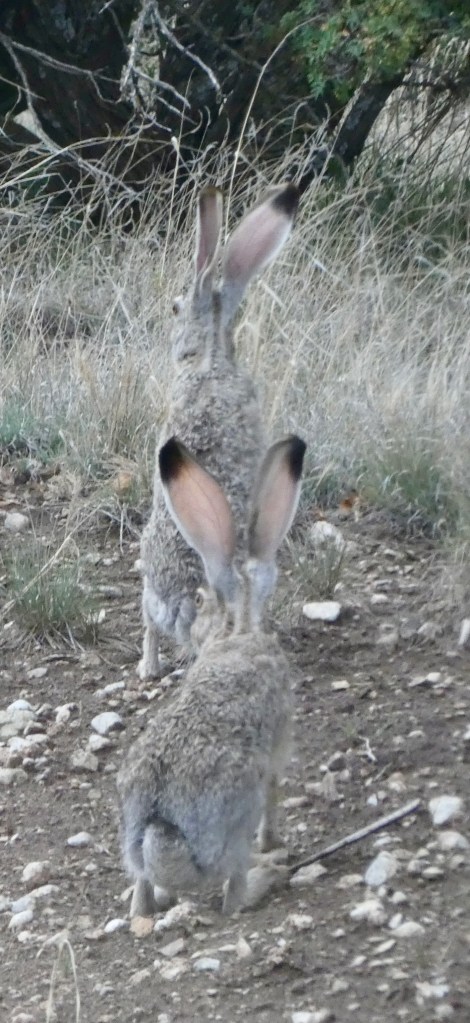


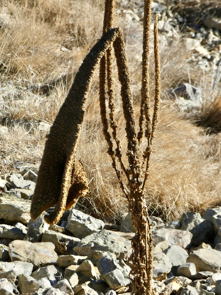
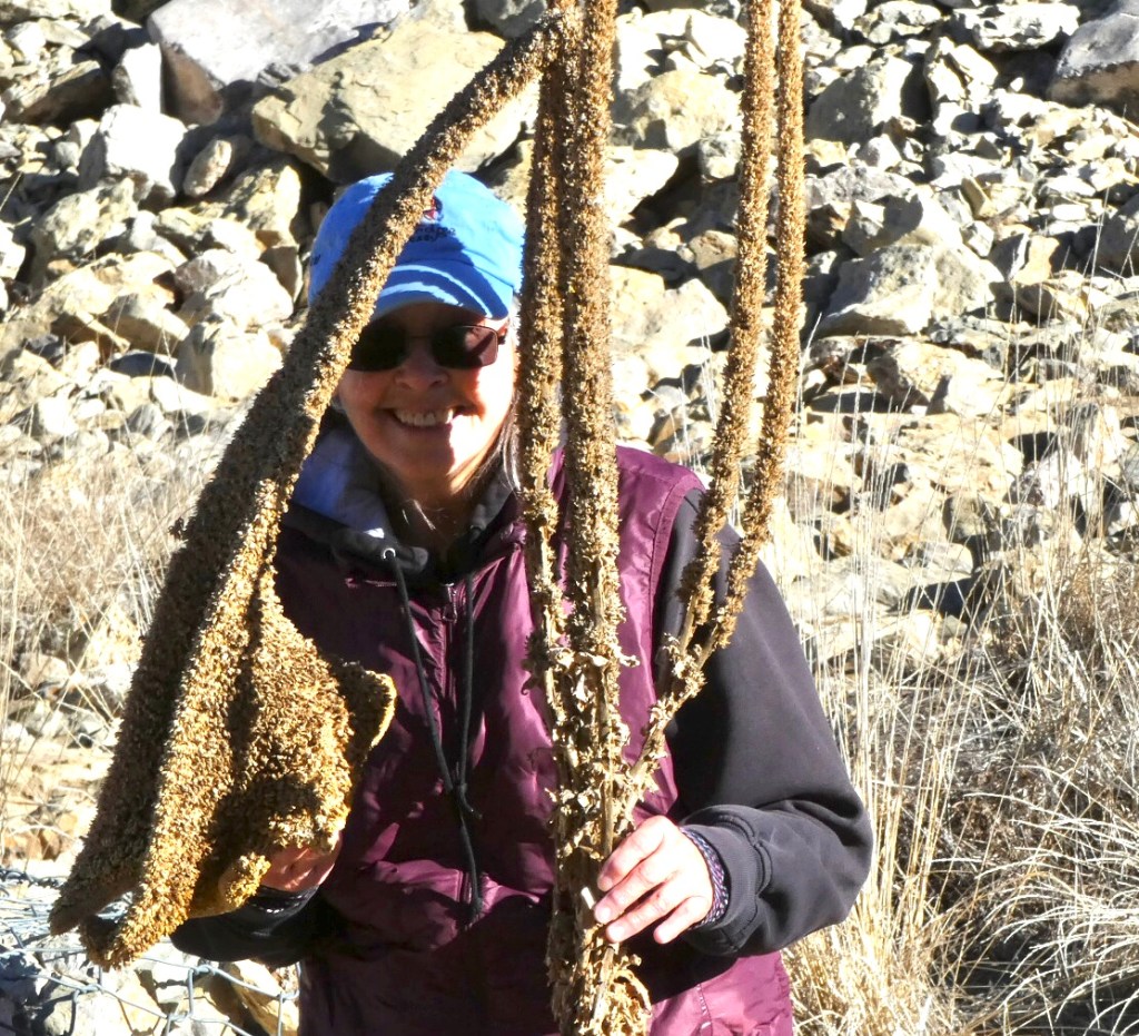
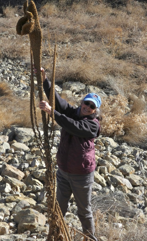
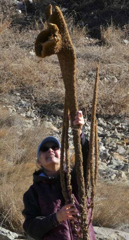
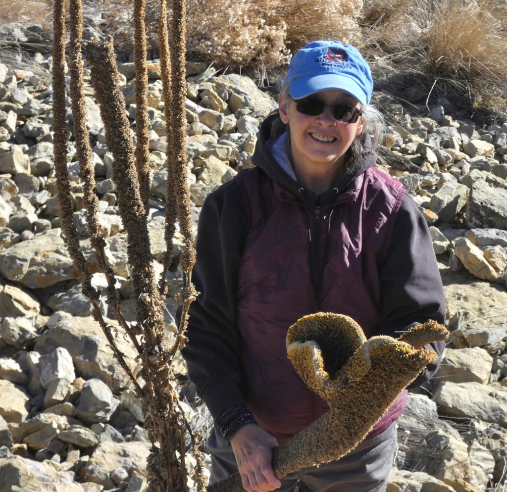
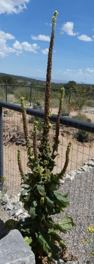
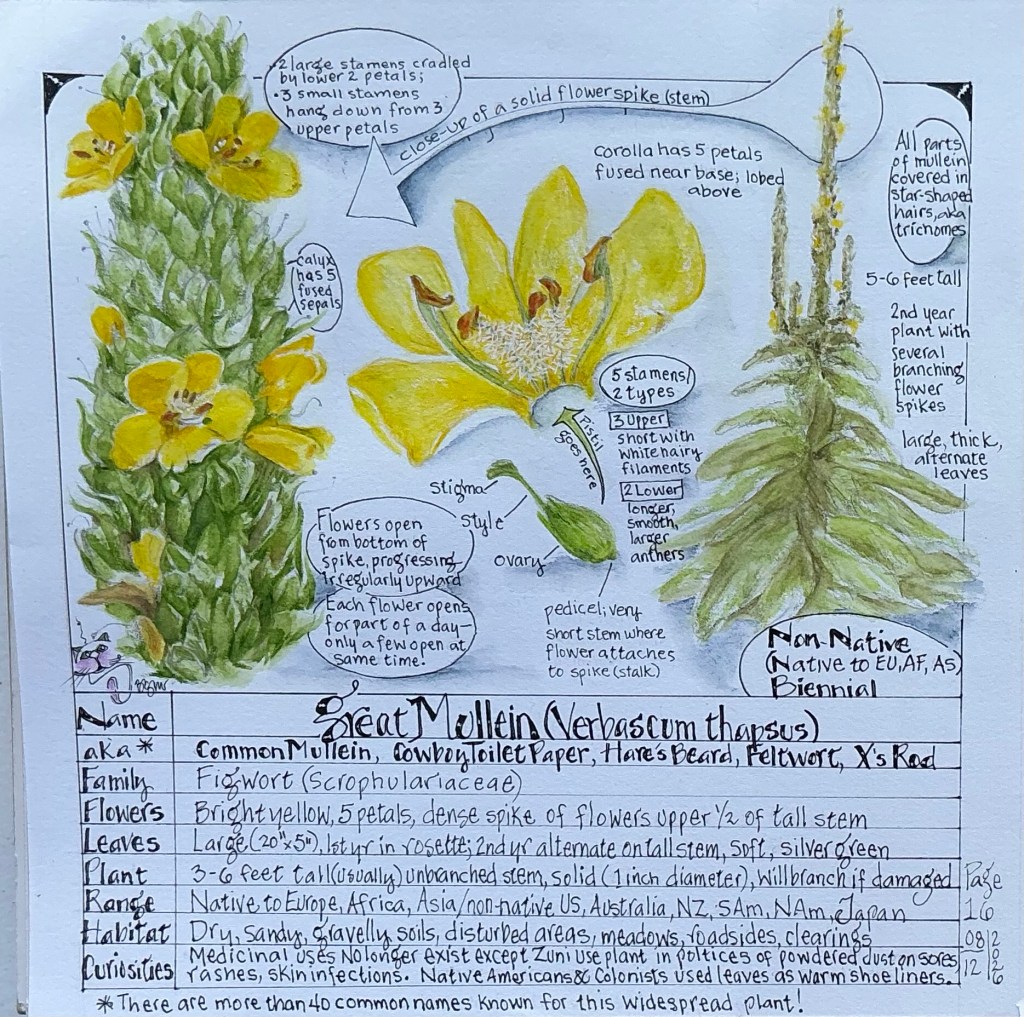
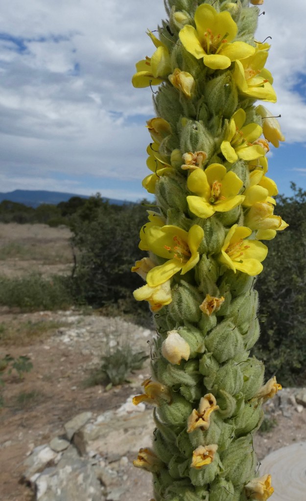
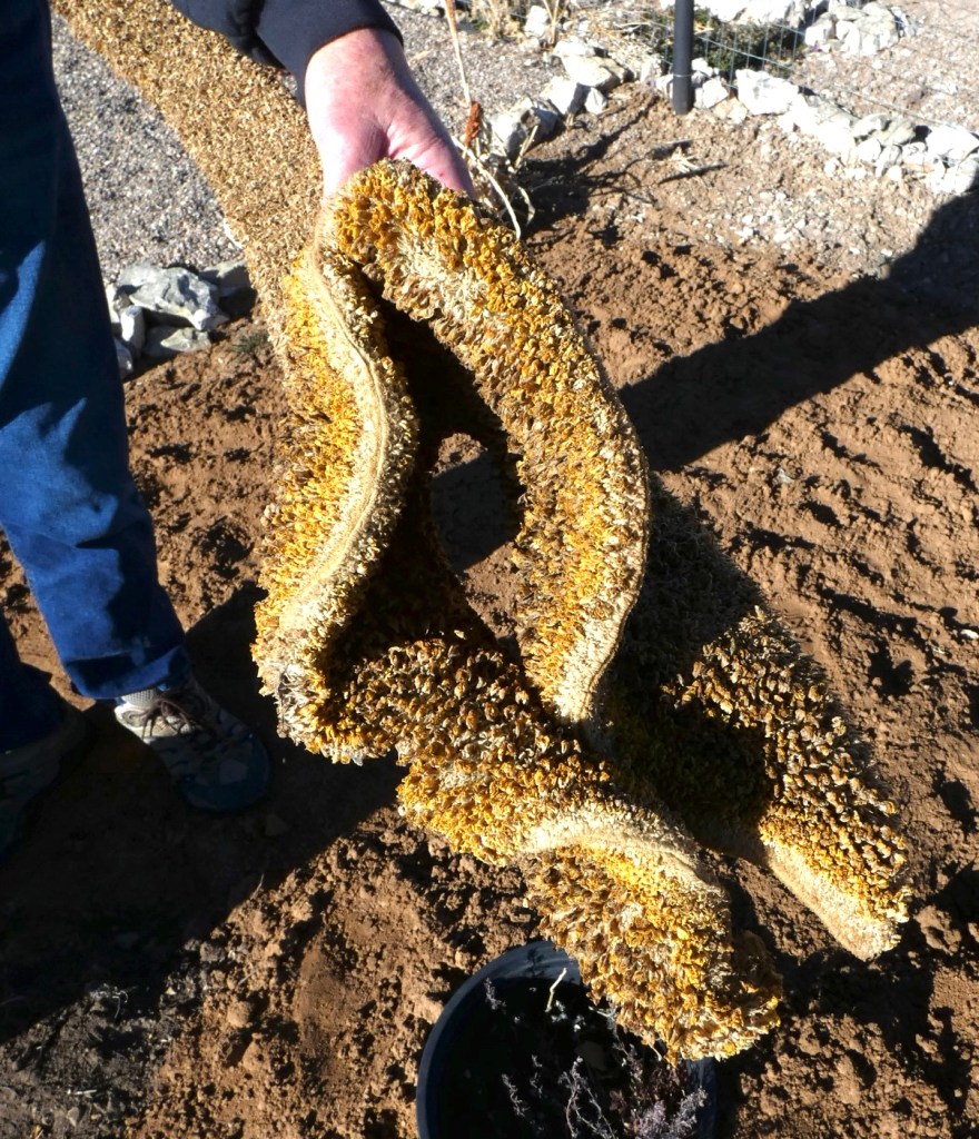
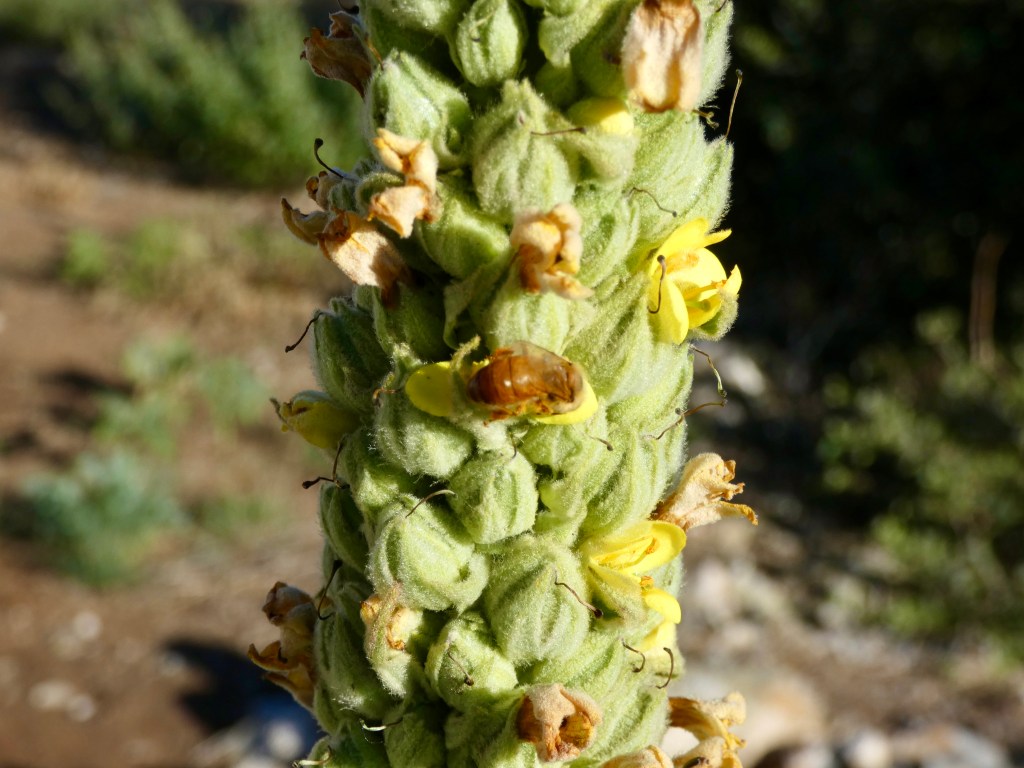
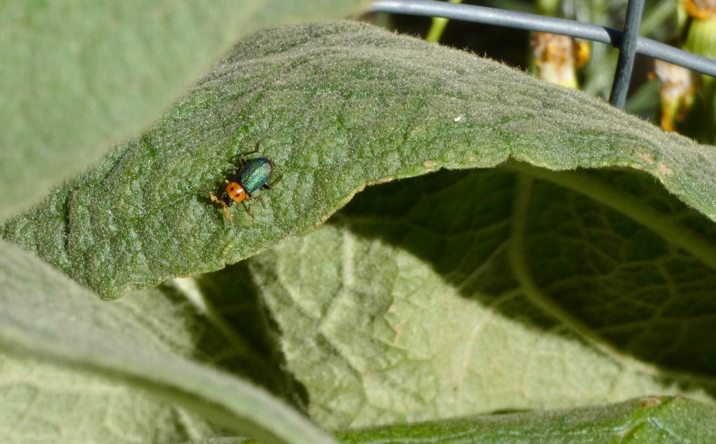
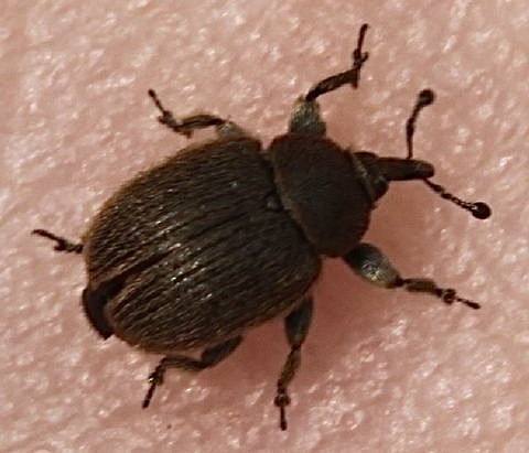
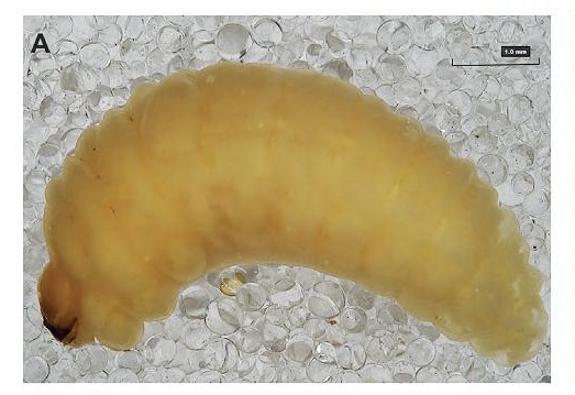
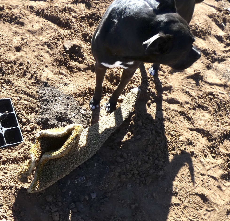


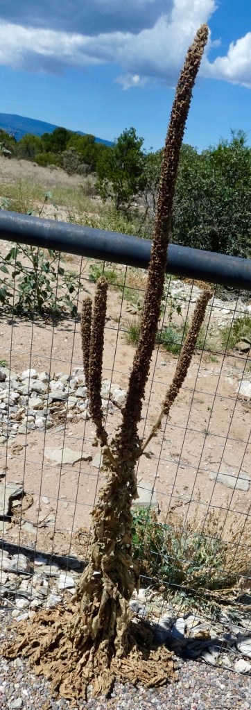
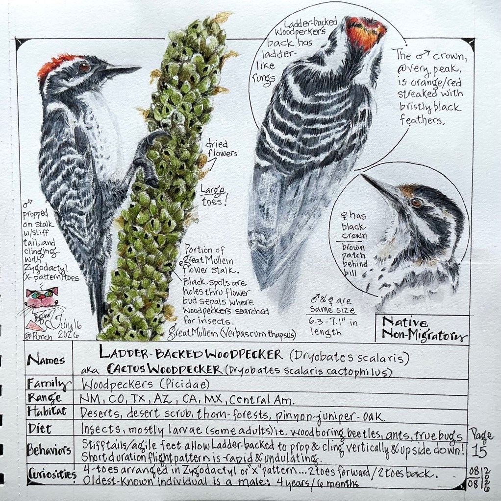
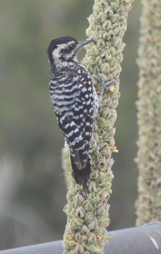
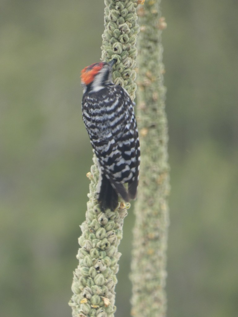
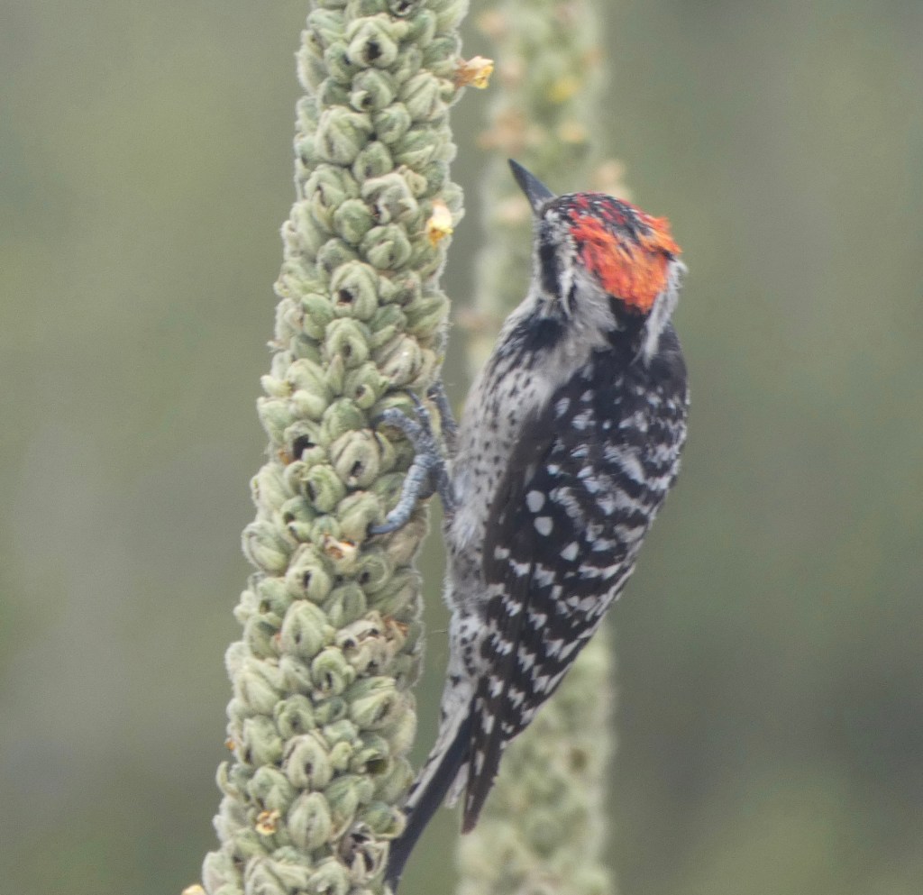
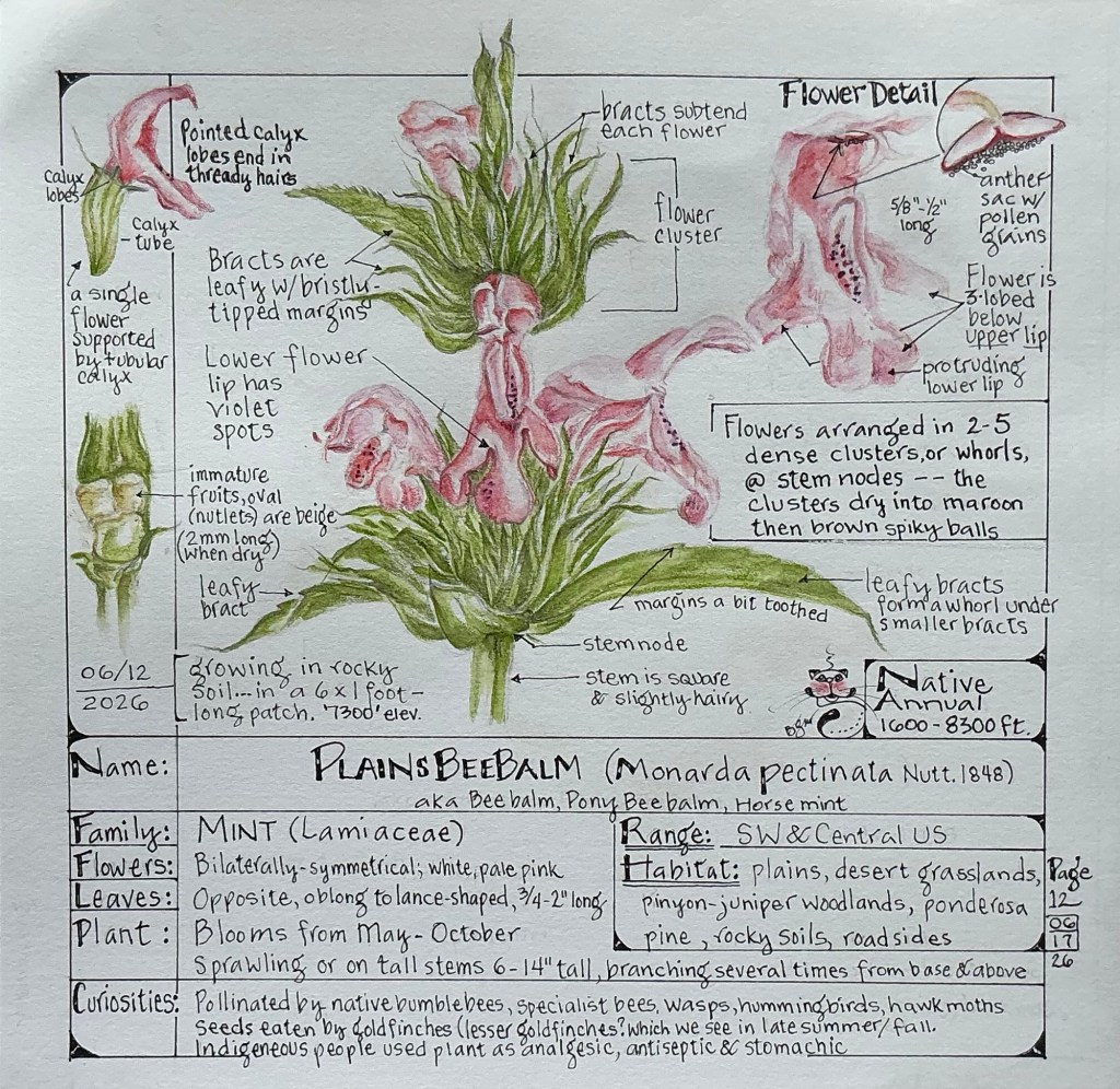
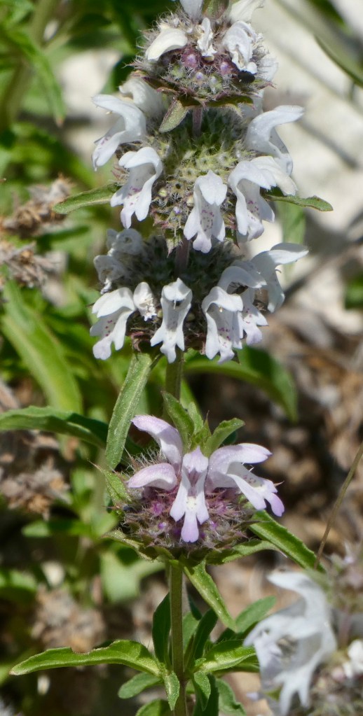
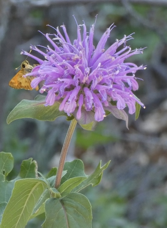
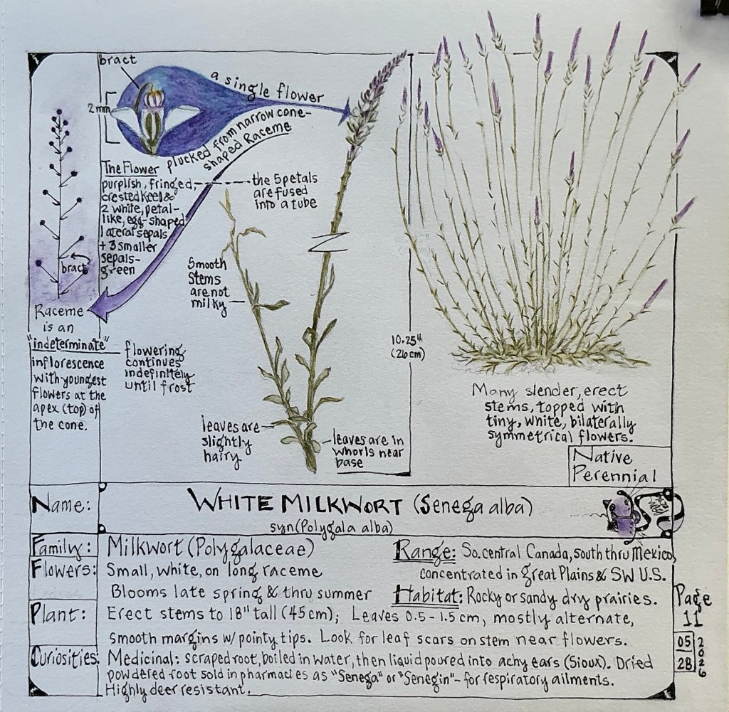
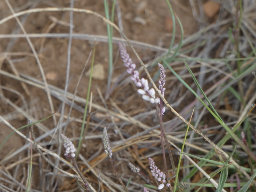
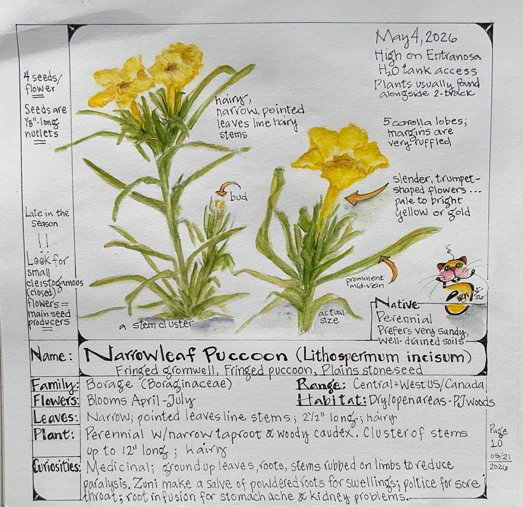
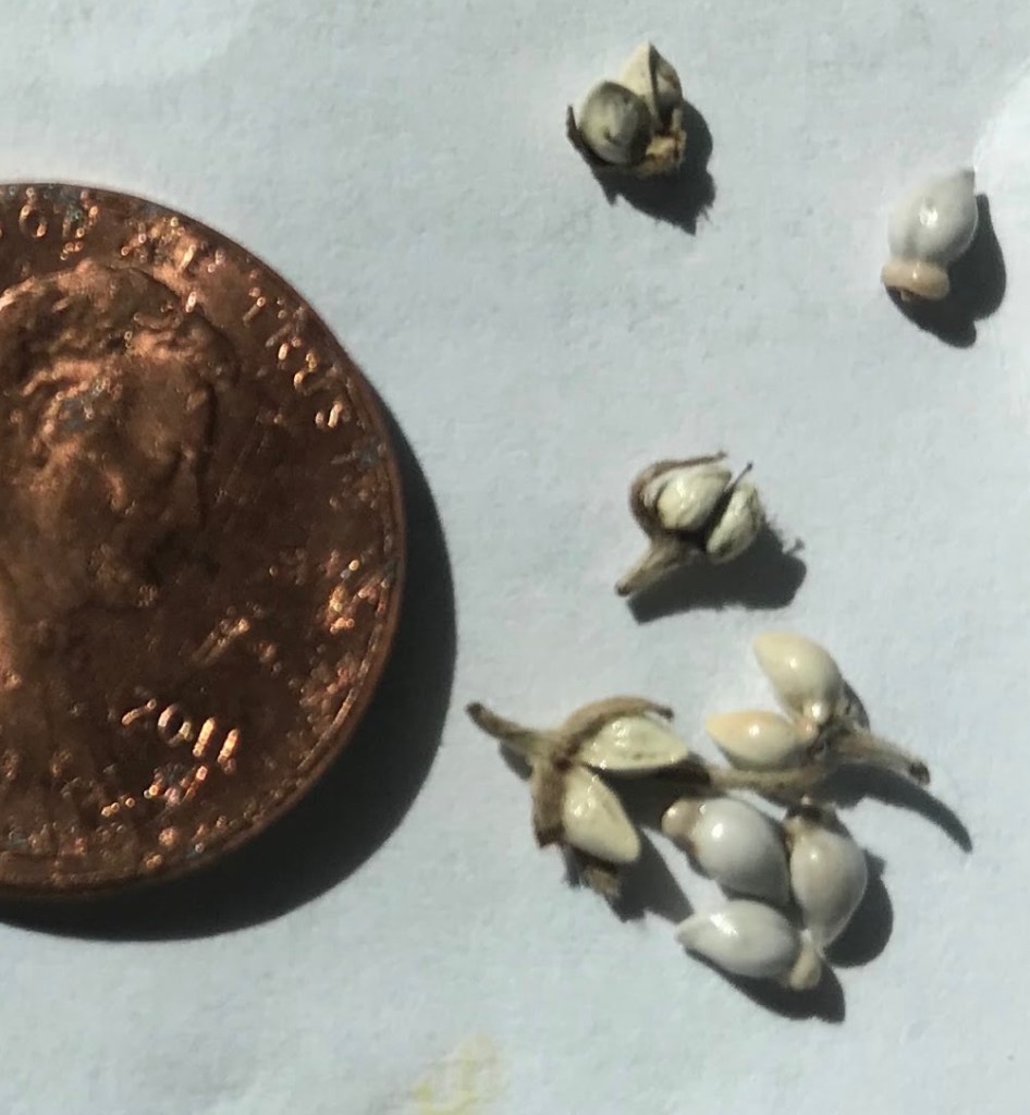
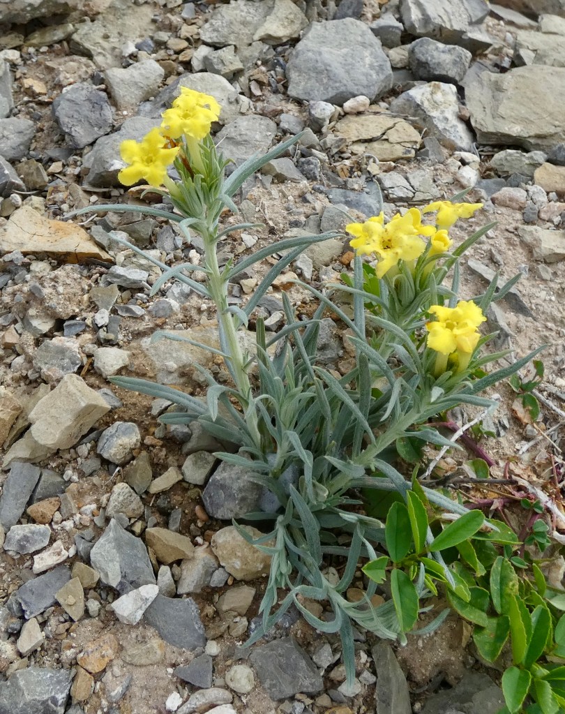
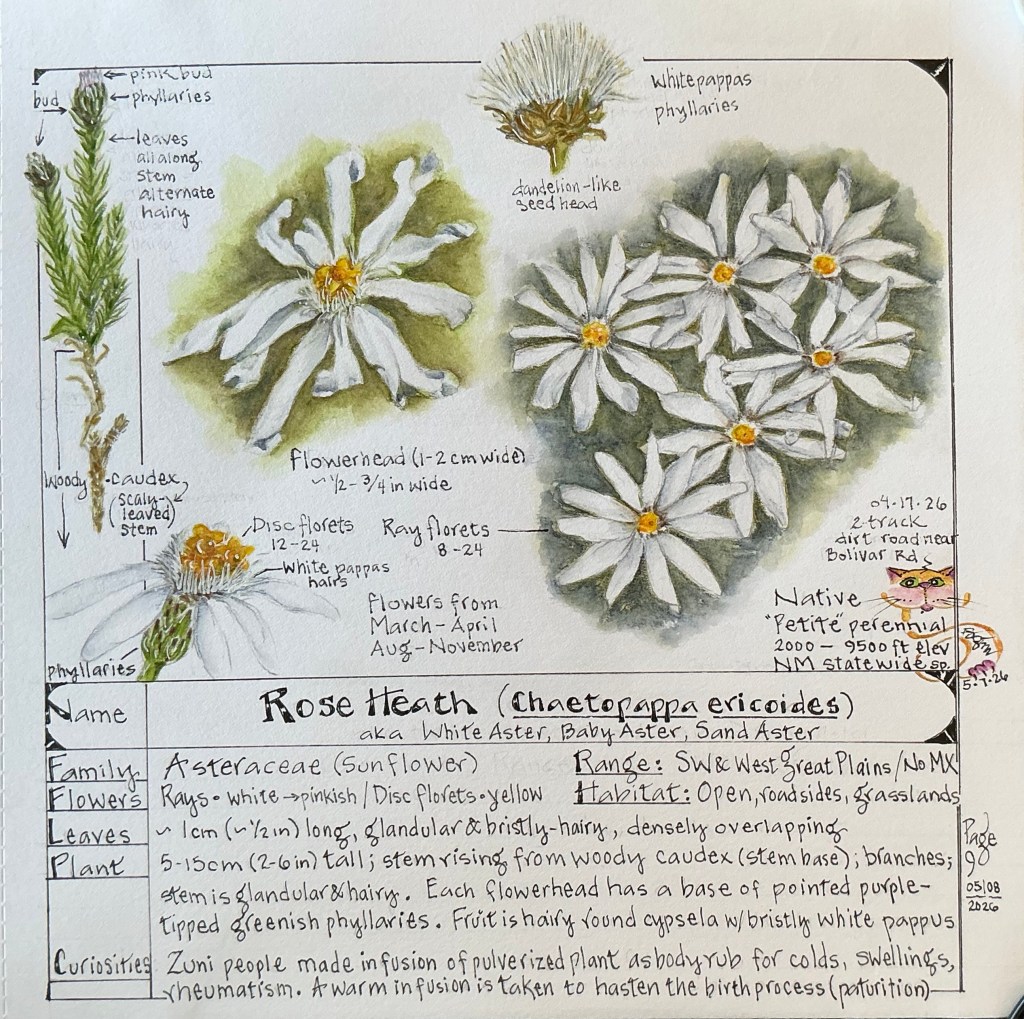
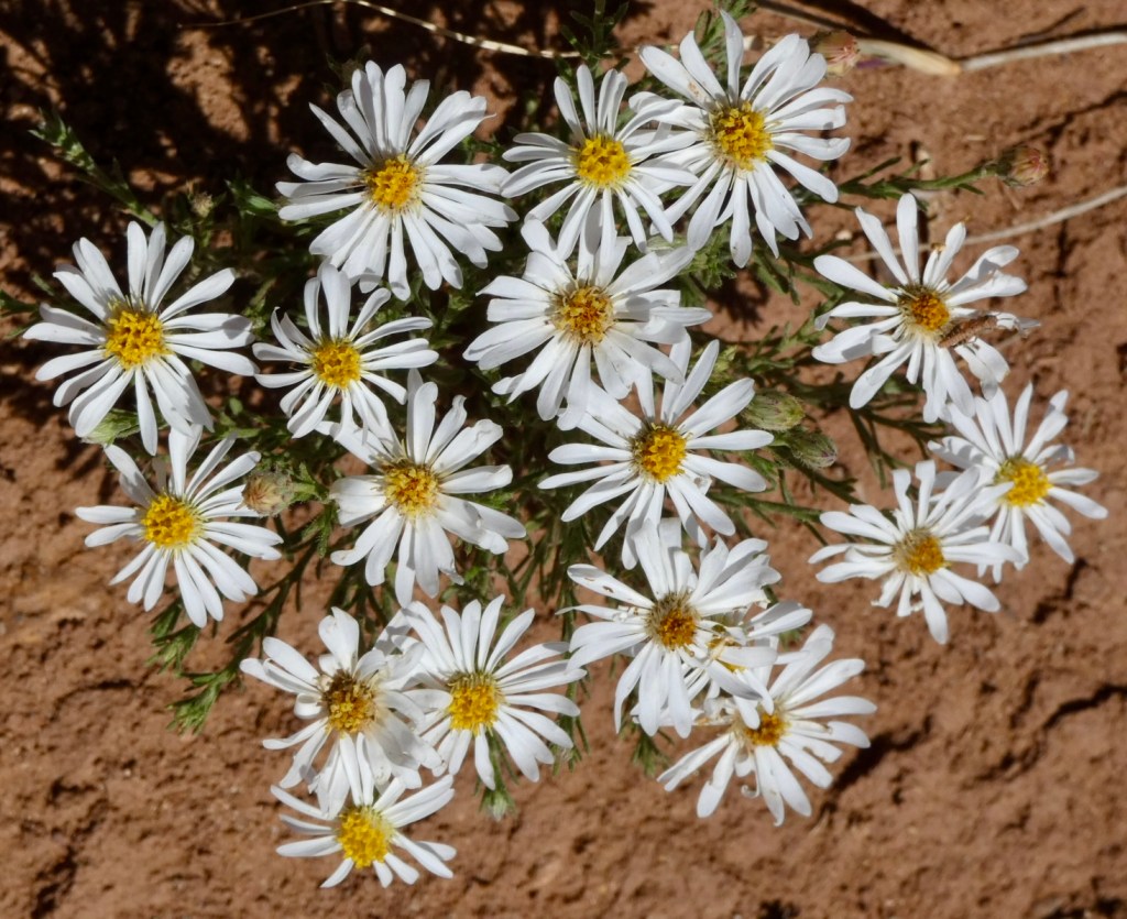
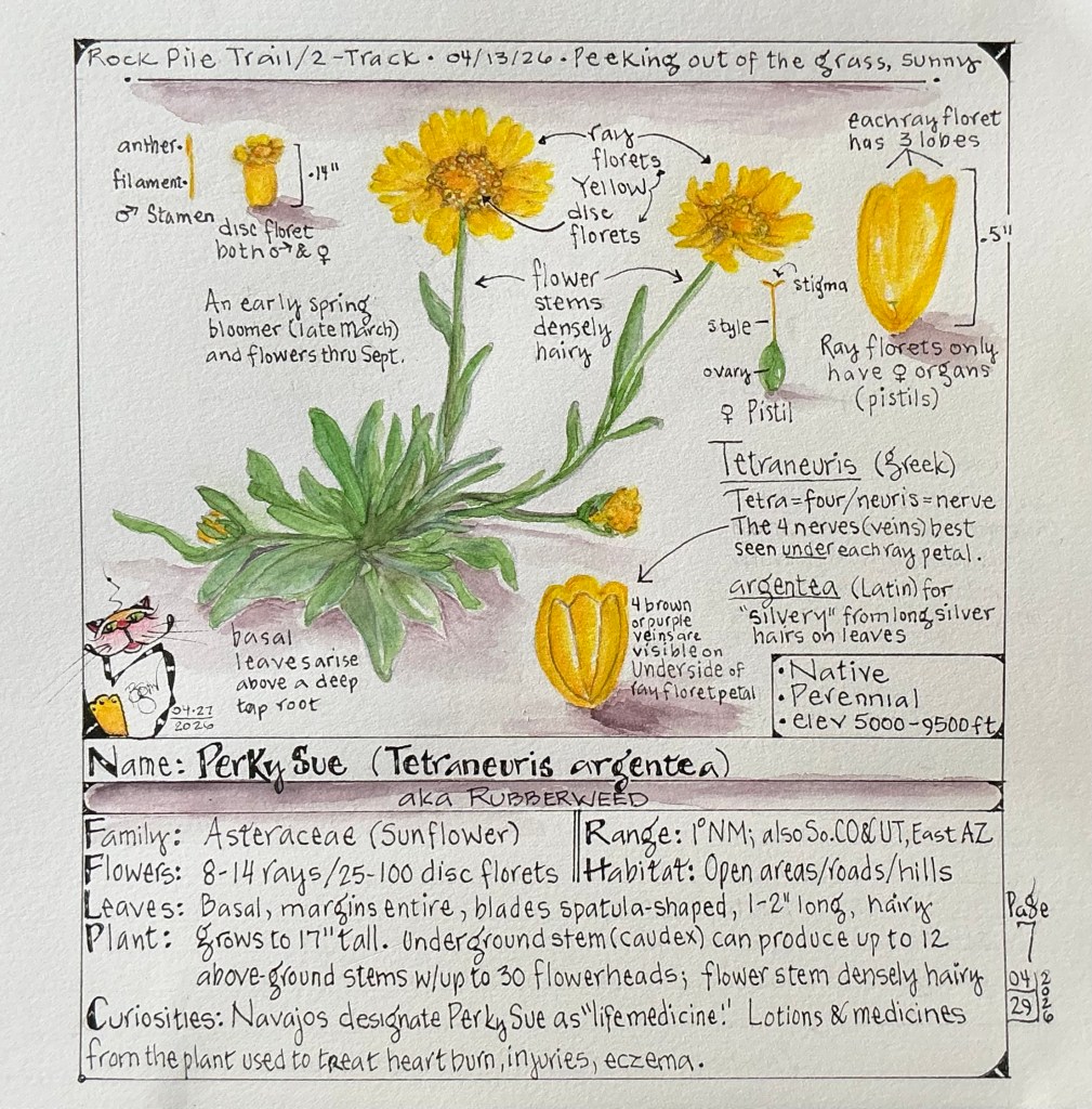
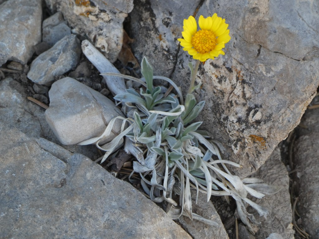
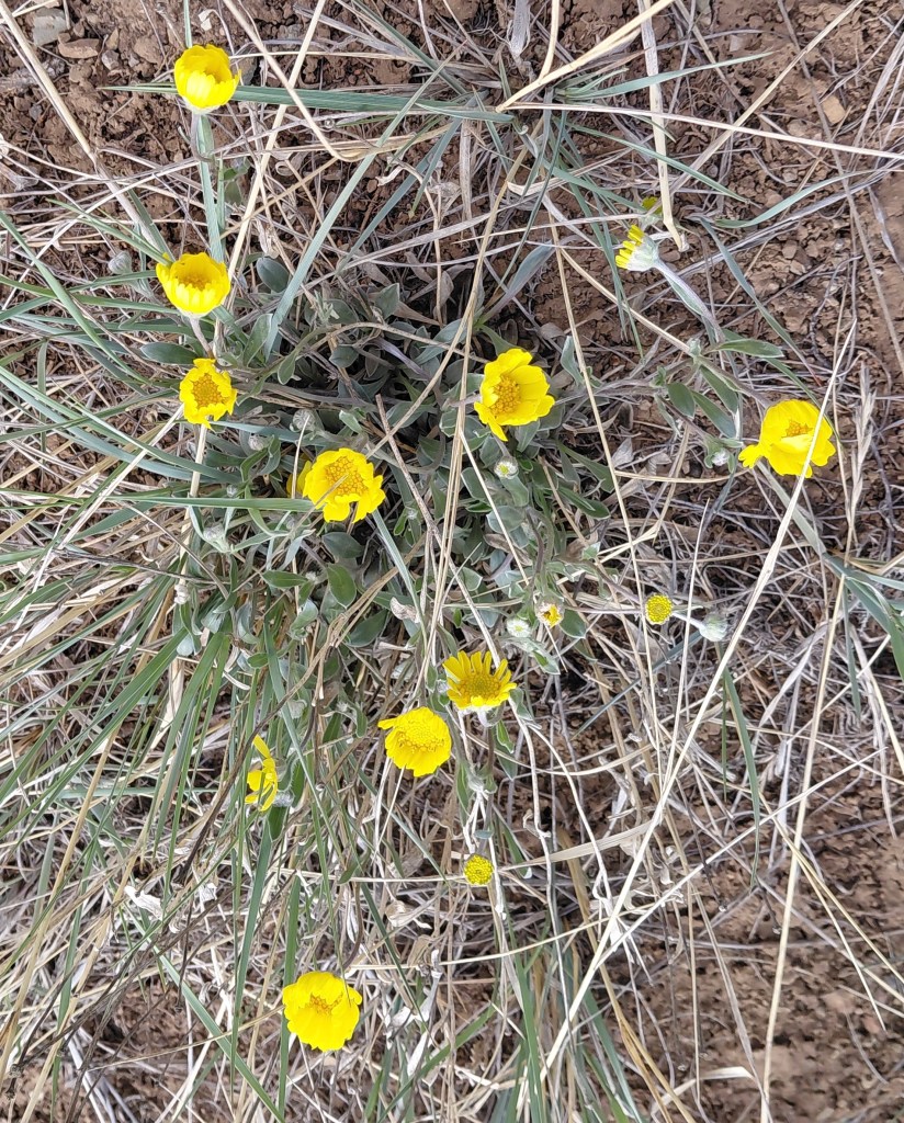
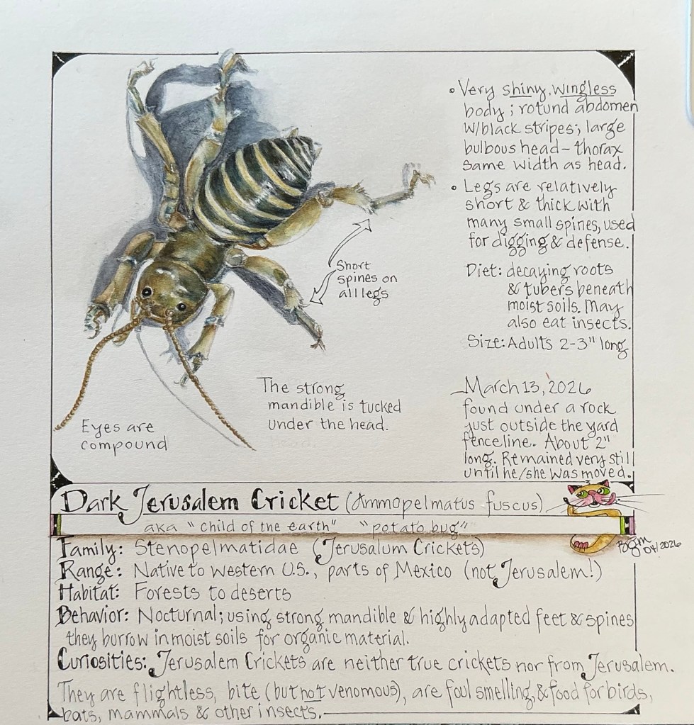
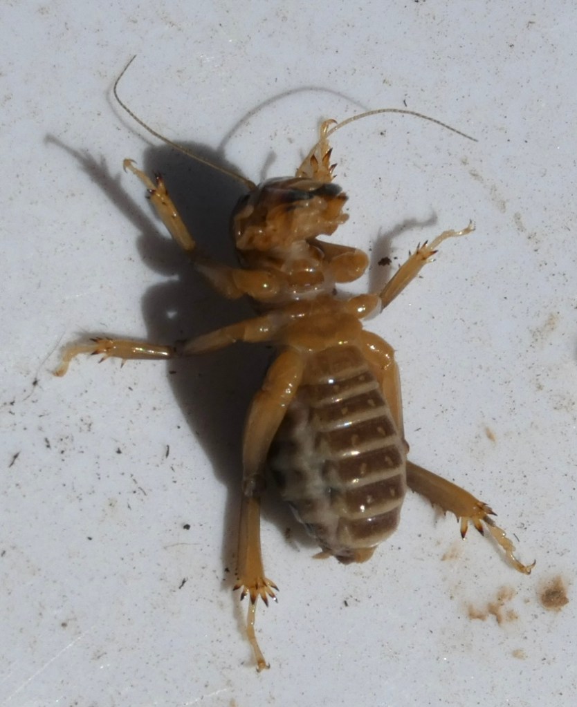
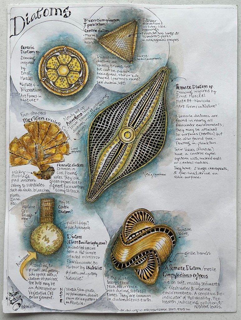
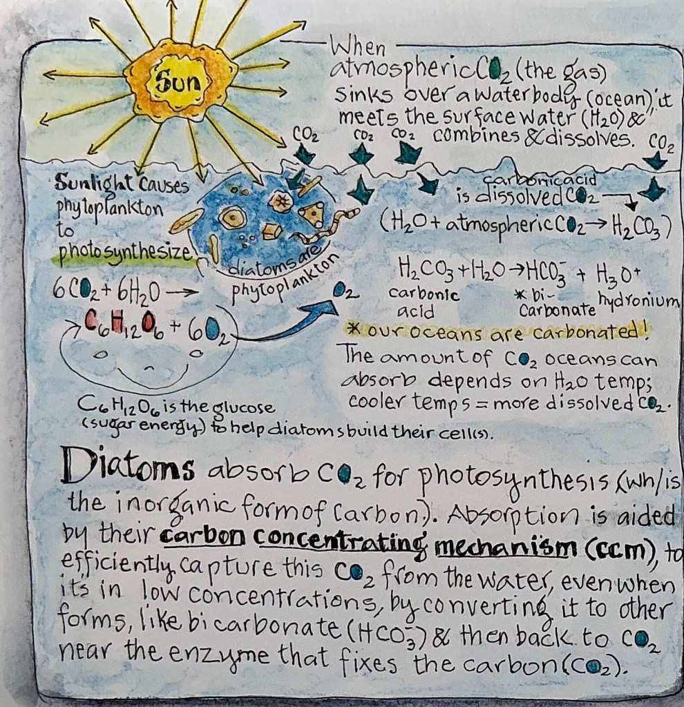
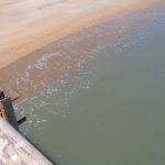
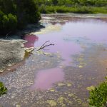
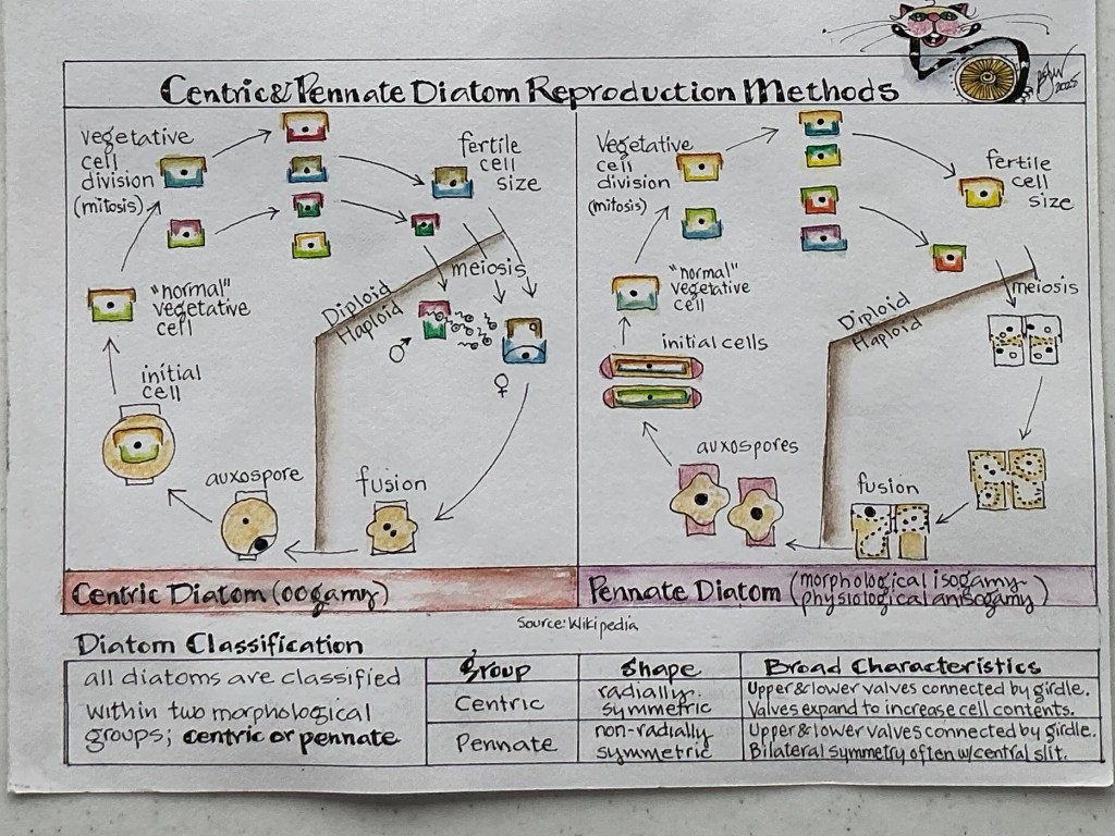
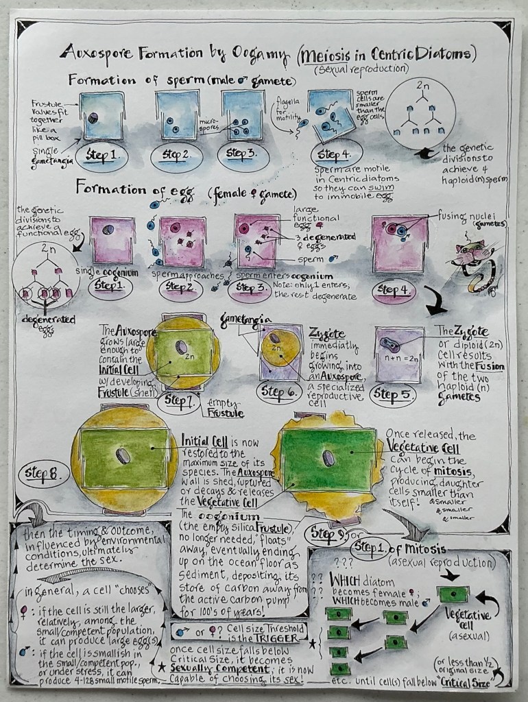
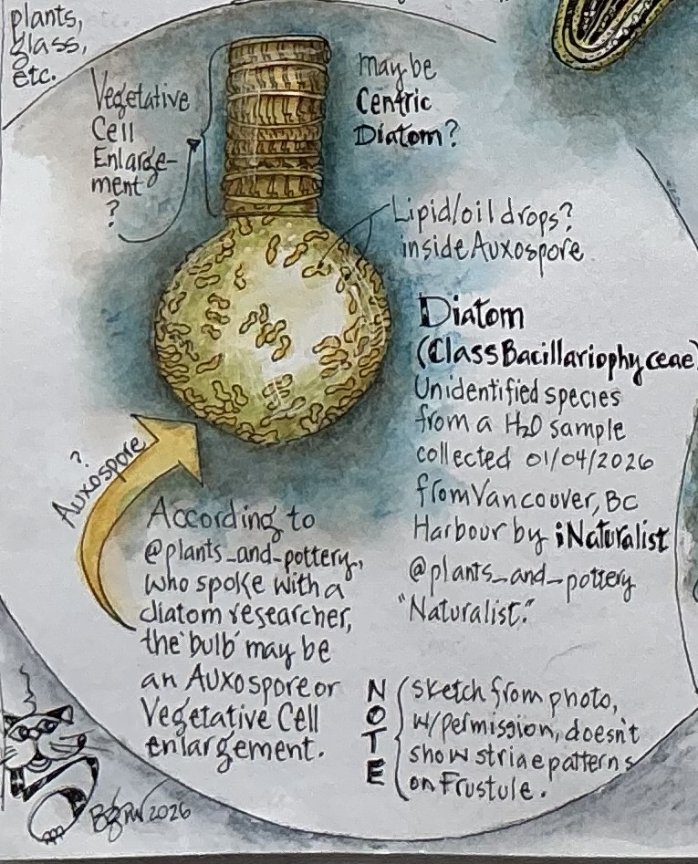
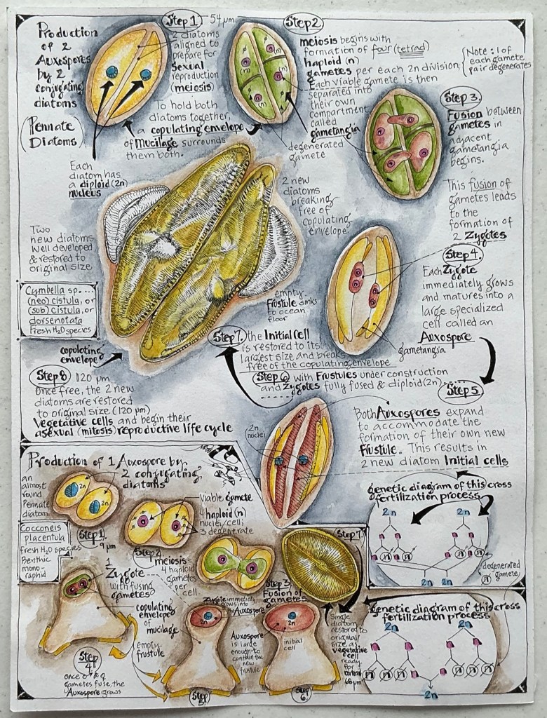
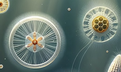
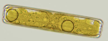
 2𝑛), forming the dominant, actively growing stage, which produces haploid (𝑛) gametes through meiosis, that fuse to restart the cycle by restoring two sets of chromosomes (
